Cervical Spondylosis Treatment in Patna
Relief from chronic neck stiffness, osteophyte bone spurs, shooting arm pain & cervicogenic headaches through precision radiofrequency ablation & non-surgical nerve care.
Understanding Cervical Spondylosis (Neck Osteoarthritis)
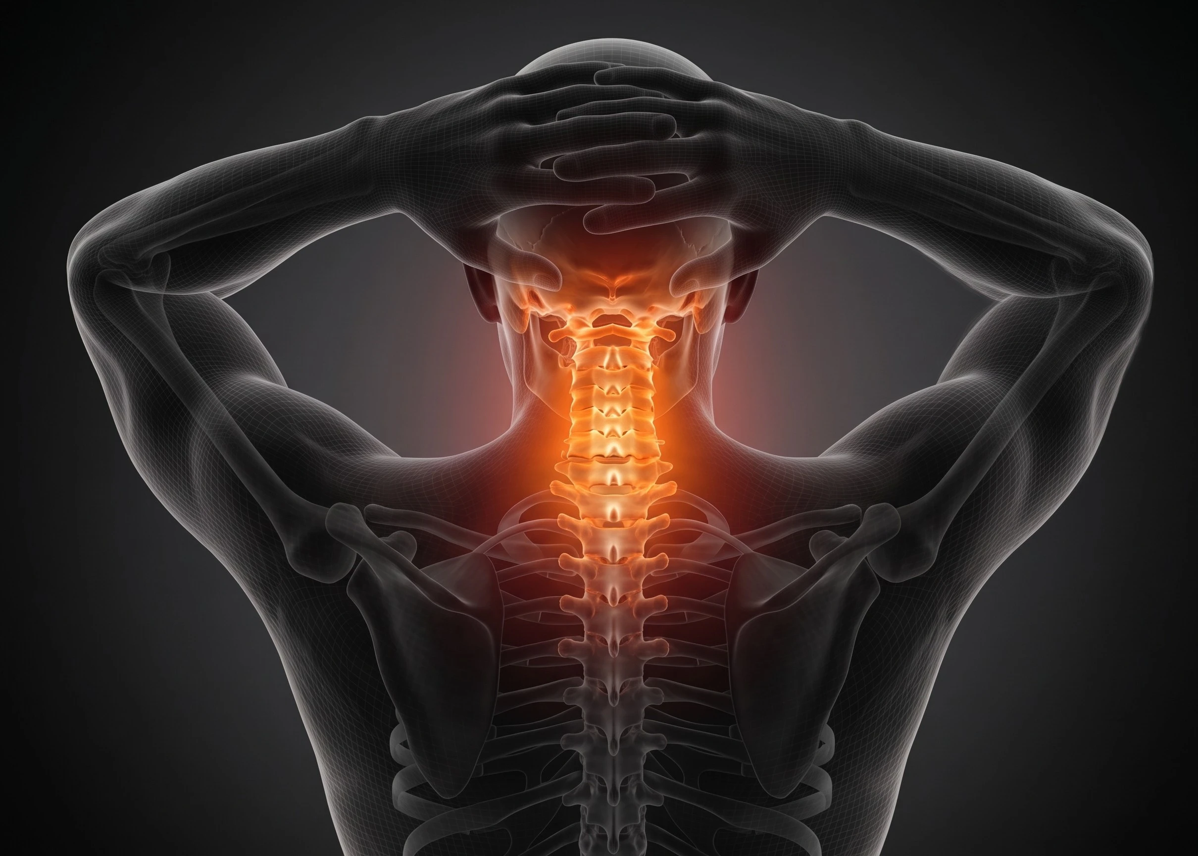
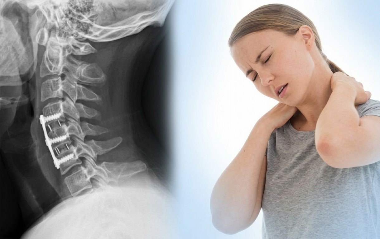
Cervical Spondylosis is a generalized medical term for age-related and posture-induced wear and tear affecting the cervical spine (C1 to C7 vertebrae). It involves progressive degeneration of the shock-absorbing intervertebral discs, the paired facet joints, uncovertebral joints (joints of Luschka), and surrounding stabilizing spinal ligaments.
As cervical discs gradually dehydrate and lose height, excess mechanical stress shifts onto the posterior facet joints, leading to cartilage erosion and localized friction. In response to this instability, the body forms extra bone overgrowths called Osteophytes (Bone Spurs).
These degenerative changes typically trigger three distinct clinical problems:
- Axial Neck Pain & Stiffness: Arthritic inflammation of facet joints and paraspinal muscle spasms restricting head rotation.
- Cervical Radiculopathy: Bone spurs and herniated disc margins narrowing exit foramina, pinching nerve roots traveling to the shoulder, arm, and hand.
- Cervicogenic Headaches: Irritation of upper cervical nerves (C1–C3) referring throbbing pain to the base of the skull, temples, and behind the eyes.
Interventional Pain Medicine pinpoints the exact arthritic joint or compressed nerve root under live C-Arm fluoroscopy, providing long-lasting non-surgical relief without open spinal fusion surgeries or metal implants.
How Cervical Spondylosis Manifests
Cervical spondylosis affects patients differently depending on whether the primary problem is in the joints, exiting nerves, or spinal cord.
1. Axial Neck Arthritis (Facet Pain)
Localized, aching neck stiffness that worsens when looking up, turning your head, or after hours of computer desk work. Associated with a grinding or cracking sound (Crepitus).
- Primary Cause: Cervical facet joint cartilage degeneration.
- Associated: Upper shoulder blade ache & occipital headache.
2. Cervical Radiculopathy (Pinched Nerve)
Sharp, electric-shock pain, burning, tingling, and numbness shooting from the neck through the shoulder, bicep/tricep, and into the fingers, caused by bone spurs or disc bulges narrowing exit canals.
- Primary Cause: Foraminal stenosis (C5, C6, C7 nerve roots).
- Associated: Weakened arm lifting, grip strength or finger drop.
3. Cervicogenic Headaches & Dizziness
Chronic unilateral throbbing pain originating at the upper neck (C1-C3) and radiating over the skull, temples, and behind the eyes. Often confused with typical migraine or tension headaches.
- Primary Cause: Upper cervical facet & occipital nerve irritation.
- Associated: Lightheadedness upon rapid neck movement.
Cervical Nerve Root Distribution & Symptoms
The specific location of your arm pain, numbness, and muscle weakness directly pinpoints which cervical spinal level is compressed.
| Cervical Spine Level | Compressed Nerve | Pain & Numbness Path | Motor Weakness / Reflex Deficit |
|---|---|---|---|
| C4 – C5 Level | C5 Nerve Root | Neck, top of shoulder (deltoid area), and upper arm | Weak shoulder abduction (difficulty lifting arm sideways); Deltoid weakness |
| C5 – C6 Level | C6 Nerve Root | Side of neck, biceps, outer forearm, thumb, and index finger | Weak bicep flexion and wrist extension; Diminished Biceps Jerk reflex |
| C6 – C7 Level | C7 Nerve Root | Back of neck, triceps, back of forearm, and middle finger | Weak triceps (difficulty pushing away) & wrist flexion; Diminished Triceps reflex |
| C7 – T1 Level | C8 Nerve Root | Inner forearm, ring finger, and little finger | Weak hand grip, dropping objects, difficulty with fine buttoning/writing tasks |
Our Diagnostic Protocol for Cervical Spondylosis
Combining thorough neurological examination with advanced imaging ensures exact identification of the arthritic pain generator.
Specialized Neck Exams
Spurling's compression test, Hoffman's reflex test, neck range of motion testing, and dermatome sensory checks.
Dynamic Cervical X-Rays
Flexion, extension, and oblique views to detect osteophyte spurs, uncovertebral arthrosis, and segmental motion instability.
High-Resolution 3T MRI
Visualizes disc bulges, foraminal stenosis, thecal sac crowding, and evaluates spinal cord integrity for myelomalacia.
Diagnostic Facet Block
Targeted micro-injection of local anesthetic over the medial branch nerves under C-Arm to confirm joint-origin pain before RFA.
Non-Surgical Interventions for Cervical Spondylosis
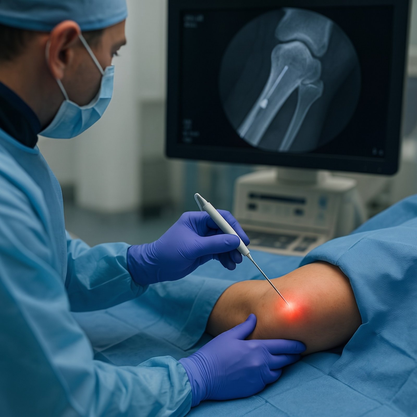
State-of-the-art daycare interventional procedures performed under real-time C-Arm fluoroscopic and ultrasound guidance by Dr. Shrutika Bhagat.
 Long-Term Relief
Cervical Facet Radiofrequency (RFA)
C-Arm guided thermal radiofrequency energy applied to the medial branch sensory nerves supplying arthritic facet joints, deactivating pain transmission and providing 12 to 24 months of sustained neck stiffness relief.
- Deactivates chronic arthritic grinding pain
- Preserves 100% natural neck flexibility
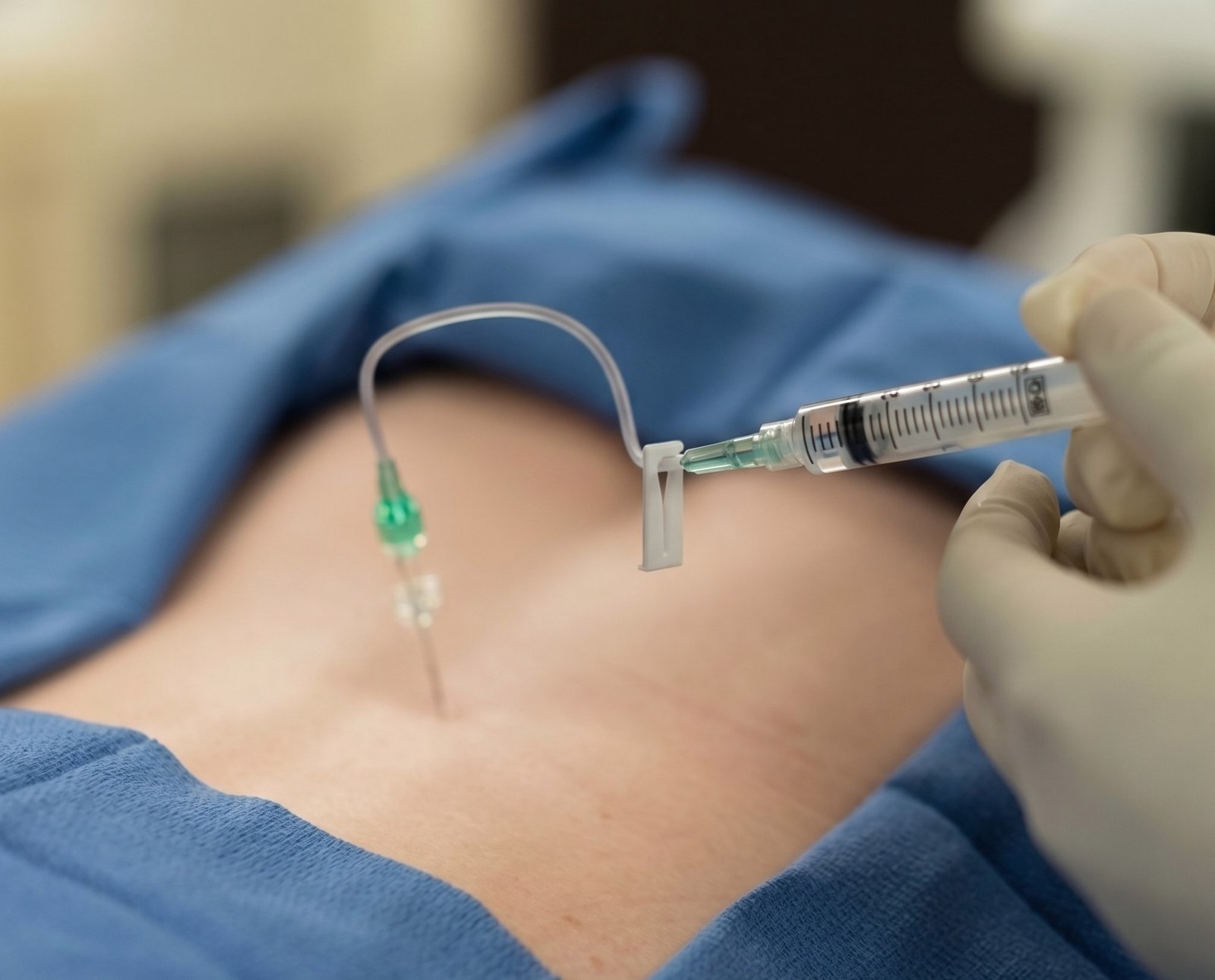 Image-Guided
Cervical Epidural Injection (TFESI)
Precision fluoroscopy-guided injection delivering concentrated anti-inflammatory medicine directly around the swollen cervical nerve root at the exit foramen, rapidly clearing radiating arm agony and finger numbness.
- Rapid relief from shooting arm & shoulder pain
- Calms swollen nerves trapped by bone spurs
 Targeted Adhesiolysis
Cervical Catheter Neuroplasty
A specialized steerable micro-catheter navigates into the cervical epidural space under live C-Arm guidance to gently dissolve dense fibrous adhesions and scar tissue surrounding entrapped cervical nerve roots.
- Mechanical release of bound nerve roots
- Ideal for severe chronic cervical spondylosis
 Headache Relief
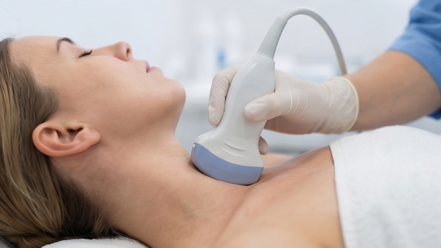
Occipital Nerve Blocks (GON/LON)
Real-time high-resolution ultrasound guidance to bathe the greater and lesser occipital nerves in anti-inflammatory medication, delivering instant relief for neck-triggered cervicogenic headaches and cranial throbbing.
- Stops severe base-of-skull and eye headaches
- Radiation-free ultrasound precision
 Biologic Repair
Cervical PRP & Facet Biotherapy
Autologous concentrated platelet-rich plasma (PRP) injected under live imaging into worn cervical facet capsules and weakened paraspinal ligaments to stimulate cellular cartilage repair and stabilize cervical motion segments.
- 100% natural, patient's own healing growth factors
- Strengthens loose, arthritic neck joints
 Keyhole Procedure
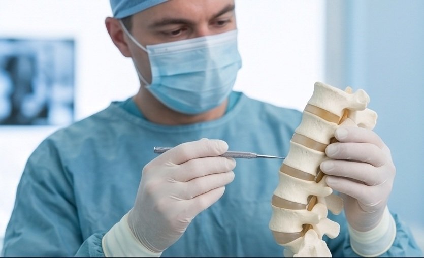
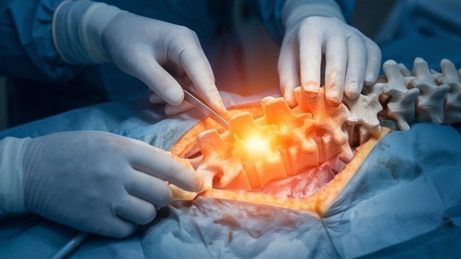
Keyhole Endoscopic Foraminotomy
Ultra-minimally invasive stitchless decompression performed through a tiny 7mm port under local anesthesia. An HD camera visualizes the narrowed exit foramen and micro-instruments widen the canal without spinal fusion.
- Zero metal plates, screws, or bone fusion
- Daycare procedure, walk home same day
Open Cervical Spine Fusion vs. Interventional Care
Why non-surgical interventional pain procedures are the preferred first-line choice for cervical spondylosis.
| Clinical Feature | Open Cervical Fusion (ACDF / Posterior) | Interventional Spine Care & RFA |
|---|---|---|
| Anesthesia Required | General Anesthesia with intubation | Local Anesthesia / Mild Conscious Sedation |
| Incision & Stitches | Large neck incision with permanent surgical scar | Micro-needle punctures (Zero stitches required) |
| Neck Motion Preservation | Permanently locks neck joints (Loss of head rotation) | 100% preserves natural neck flexibility and motion |
| Metal Implants & Hardware | Titanium plates, screws, and interbody cages | Zero metal implants or artificial hardware inserted |
| Adjacent Segment Disease Risk | High risk (Stress shifts to adjoining neck levels) | Zero adjacent segment degeneration risk |
| Hospitalization & Recovery | 3 to 5 days hospital stay; 2-3 months recovery collar | Daycare: Discharged in 2 hours; work in 24-48 hours |

Dr. Shrutika Bhagat
MBBS, MD, FIRA, FIAPM | Interventional Pain & Spine Endoscopy
Dr. Shrutika Bhagat brings super-specialized training in full-endoscopic spine surgery and image-guided interventional pain management, having successfully treated thousands of patients with severe back pain, disc prolapse, sciatica, nerve compression, and joint arthritis in Patna.
Cervical Spondylosis FAQs
Clear answers regarding neck arthritis, facet radiofrequency ablation, pillows, and non-surgical recovery.
Don't Let Chronic Pain Control Your Life.
Take the first step toward understanding your pain and exploring modern, evidence-based minimally invasive relief with Dr. Shrutika Bhagat.