Endoscopic Adhesiolysis in Patna
Direct camera visualization and gentle mechanical release of epidural scar tissue (fibrosis) for Failed Back Surgery Syndrome (FBSS) & chronic radiculopathy without re-operation.
Dissolve Post-Surgical Scar Tissue & Free Trapped Nerves
After open spine surgery (laminectomy, discectomy, or spinal fusion), up to 30% to 40% of patients develop dense epidural fibrous scar tissue (Peridural Fibrosis). This thick biological scar wraps tightly around spinal nerve roots, tethering them to the spinal canal wall and depriving them of blood flow, producing relentless leg pain and stiffness known as Failed Back Surgery Syndrome (FBSS).
Performing revision open spine surgery carries high failure rates because cutting scar tissue simply triggers more scar formation. Endoscopic Adhesiolysis (Epiduroscopy) is a breakthrough pinhole intervention performed through the natural sacral hiatus opening at the tailbone.
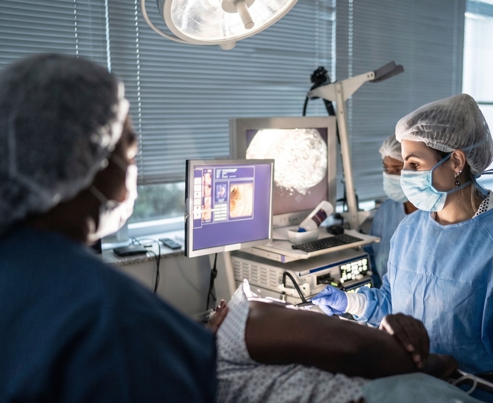
Using a steerable flexible micro-video endoscope under live fluoroscopic guidance, Dr. Shrutika Bhagat directly visualizes the inflamed, scarred nerve root. The micro-catheter delicately separates adhesions mechanically while delivering targeted enzymatic solutions (Hyaluronidase, targeted anti-inflammatories, and hypertonic wash) to dissolve fibrosis and restore smooth nerve sliding.
Who is a Candidate for Endoscopic Adhesiolysis?
Indicated for patients with persistent or recurrent spine and leg pain following prior surgeries or chronic epidural inflammation.
Failed Back Surgery Syndrome (FBSS)
Persistent or worsening back and leg pain months or years after lumbar laminectomy, microdiscectomy, or spinal fusion surgery.
Epidural Scar Fibrosis
Post-operative fibrotic bands tethering nerve roots, visible on contrast-enhanced 3T MRI as enhancing peridural soft tissue adhesions.
Chronic Refractory Radiculopathy
Constant burning, tingling, and electrical shocks radiating down both legs that failed to respond to standard epidural steroid injections.
Patients Ineligible for Revision Surgery
Patients advised that further open surgery has high risk of paralysis or complications, seeking a safe, non-cutting outpatient alternative.
Multi-Level Foraminal Adhesions
Extensive scar tissue bridging across multiple lumbar levels (L3-L4, L4-L5, L5-S1) navigated cleanly through a single sacral entry point.
Elderly Patients with Co-Morbidities
Older patients with diabetes or cardiac conditions who need definitive nerve freeing without general anesthesia or hospital stays.
Endoscopic Adhesiolysis vs. Revision Open Surgery
| Clinical Parameter | Endoscopic Adhesiolysis (Epiduroscopy) | Revision Open Spine Surgery |
|---|---|---|
| Incision & Tissue Cutting | Pinhole needle puncture via sacral hiatus | Major surgical incision cutting prior scar bed |
| New Scar Formation Risk | Minimal (Fluid-based atraumatic wash) | High (Surgical trauma triggers more fibrosis) |
| Visualization Quality | Direct 3D Video Endoscope inside epidural space | Obscured by previous surgical bleeding & scar |
| Anesthesia Type | Local Anesthesia + Conscious Sedation | General Anesthesia Required |
| Dural Tear / Nerve Damage Risk | Very low (Direct optical camera guidance) | Significantly elevated due to frozen scar planes |
| Hospital Stay | Daycare: Walk in 2 hrs; Home same day | 4 to 7 days hospital stay |
How Endoscopic Adhesiolysis is Performed
Epidurogram & Contrast Mapping
Under live C-Arm fluoroscopy, non-ionic radio-contrast is injected to identify the exact "filling defects" where scar tissue blocks medicine flow.
Sacral Hiatus Pinhole Entry
A gentle local anesthetic numbs the tailbone area. A steerable micro-catheter enters through the natural sacral canal without cutting bone.
Video Endoscope Navigation
The flexible micro-camera transmits high-definition live video of the epidural space, distinguishing healthy dura from inflamed fibrotic bands.
Mechanical Adhesion Lysis
The steerable tip gently navigates around nerve roots, peeling away fibrous bands and releasing the trapped, tethered spinal nerves.
Targeted Enzymatic Infusion
High-purity Hyaluronidase, hypertonic saline, and localized anti-inflammatory solutions are delivered directly to wash away microscopic scar tissue.
Band-Aid & Same-Day Discharge
The micro-catheter is withdrawn. A small Band-Aid is applied at the tailbone and the patient walks comfortably within 2 hours.
Adhesiolysis Techniques Offered in Patna
Direct Epiduroscopy
Micro-fiberoptic camera guidance inside the spinal canal for exact visual confirmation of scar lysis.
Racz Catheter Neurolysis
Specialized spring-wound fluoroscopic catheter designed specifically to navigate into anterior foraminal scar recesses.
Targeted Hyaluronidase Wash
Medical-grade enzyme protocol that breaks down hyaluronic acid mucopolysaccharides in fibrous scar tissue.
Epidural PRF Neuromodulation
High-frequency pulsed radiofrequency delivered through the catheter to calm chronified hypersensitive dorsal root ganglia.
Common Questions on Endoscopic Adhesiolysis
Why Choose Dr. Shrutika for Endoscopic Epidural Adhesiolysis in Patna
- Direct Endoscopic Camera Vision: Micro-endoscopes clearly visualize internal epidural scar tissue, differentiating healthy neural roots from dense post-surgical fibrosis.
- Breakthrough for Failed Back Surgery (FBSS): Provides definitive relief for patients struggling with persistent pain after prior laminectomy or fusion without repeat open surgery.
- Targeted Anti-Adhesive & Enzyme Infusion: Combines gentle mechanical separation with localized anti-fibrotic enzymes and anti-inflammatory washes to prevent recurrent adhesions.
- Safe Sacral Hiatus Approach: Natural anatomical entry with zero bone removal, minimal tissue trauma, and zero risk to spinal structural stability.
- Quick Outpatient Recovery: Comfortable procedure performed under local anesthesia with rapid same-day daycare discharge.
- Expertise in Complex Spine Cases: Specialized experience treating severe chronic nerve entrapment and refractory post-operative spinal pain in Bihar.

Don't Let Chronic Pain Control Your Life.
Take the first step toward understanding your pain and exploring modern, evidence-based minimally invasive relief with Dr. Shrutika Bhagat.