Endoscopic Cervical Decompression in Patna
Stitchless 6mm keyhole posterior cervical foraminotomy for pinched neck nerves, disc herniations & radiating arm pain. 100% natural neck mobility without metal fusion plates or screws.
Preserve Neck Motion & Eliminate Radiating Arm Pain
Posterior Endoscopic Cervical Decompression (PECD / Foraminotomy) is the world's most advanced minimally invasive surgical procedure for cervical disc herniations and foraminal nerve root compression in the neck.
Traditional neck surgery (such as ACDF - Anterior Cervical Discectomy & Fusion) requires cutting through the front of the throat, retracting the vocal cords and esophagus, removing the entire intervertebral disc, and fusing adjacent vertebrae with titanium plates and screws. In contrast, endoscopic cervical decompression is performed from the back of the neck through a single 6 to 7 millimeter pencil-thin cannula.
Under continuous saline irrigation and 4K ultra-high definition camera magnification, Dr. Shrutika Bhagat gently unroofs the narrowed neural foramen (the exit tunnel of the pinched nerve) and removes only the extruded disc fragment. 100% of your natural cervical disc, vertebral bones, and head rotation mobility remain intact—eliminating the need for artificial disc replacements or permanent metal fusion implants.
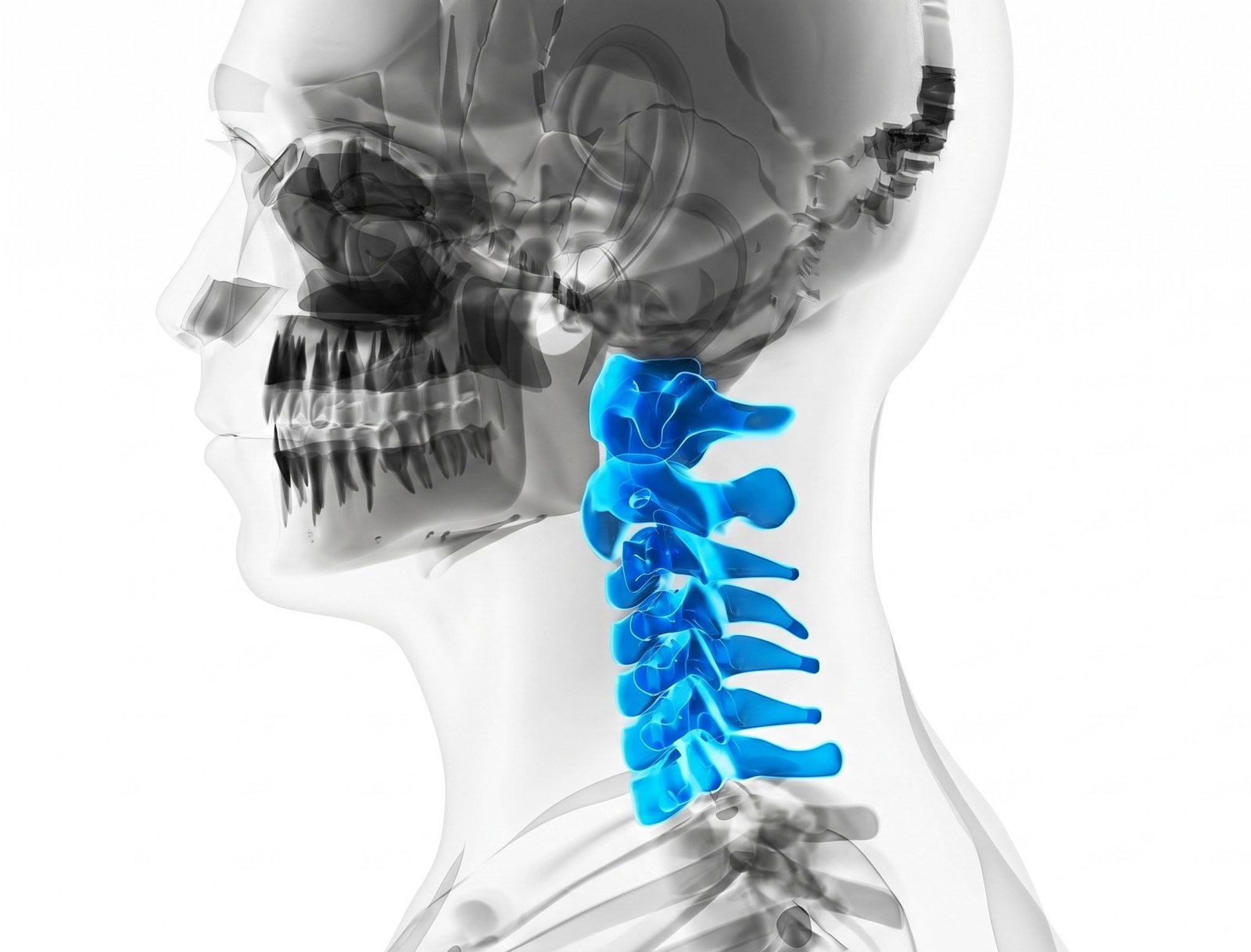
Who is a Candidate for Endoscopic Cervical Decompression?
Indicated for patients with cervical nerve root compression causing severe neck, shoulder, arm, or hand symptoms failing conservative care.
Cervical Radiculopathy & Arm Pain
Sharp, electric-shock pain shooting from the neck across the shoulder blade, radiating down the arm, forearm, thumb, or fingers (C5, C6, C7, C8 roots).
Hand Numbness & Grip Weakness
Tingling in the fingers (pins and needles) or motor weakness such as dropping cups, difficulty buttoning shirts, or loss of forearm and bicep strength.
Cervical Foraminal Stenosis
Bone spurs (osteophytes) and thickened uncinate processes pinching the exiting spinal nerve root inside the neural exit tunnel.
Patients Wanting to Avoid Neck Fusion (ACDF)
Active individuals, drivers, athletes, and professionals who refuse neck fusion plates that permanently lock neck movement and cause adjacent-segment degeneration.
Adjacent Segment Disease (ASD)
New disc herniation developing directly above or below a previous anterior cervical fusion surgery, safely decompressed through posterior keyhole routes.
Elderly & Medically High-Risk Patients
Seniors with diabetes, cardiac disease, or osteoporosis who cannot withstand open anterior surgical trauma, major bleeding, or long general anesthesia.
Endoscopic Cervical Decompression vs. Traditional ACDF Fusion
| Clinical Parameter | Endoscopic Cervical Decompression | Traditional Open ACDF Neck Fusion |
|---|---|---|
| Incision Location & Size | Posterior Neck: 6 - 7 mm (Band-Aid) | Anterior Throat: 40 - 60 mm (Visible Scar) |
| Neck Motion & Flexibility | 100% Preserved (Full Head Rotation) | Permanently Locked / Stiff Neck Segment |
| Hardware & Implants | Zero (No Metal Plates, Screws, or Cages) | Titanium Plates, Screws & Interbody Cage |
| Throat / Voice Complications | Zero (Posterior approach avoids throat) | Risk of hoarse voice & swallowing pain (Dysphagia) |
| Adjacent Segment Disease (ASD) | Virtually Zero (No fusion stress on nearby discs) | Up to 25% require 2nd surgery within 5 years |
| Hospital Stay & Collar Wear | Same-Day Discharge; Soft collar 3–5 days | 3 to 5 days hospital stay; Hard collar 6–8 weeks |
How Endoscopic Cervical Decompression is Performed
3T Cervical MRI & CT Mapping
Millimeter-precise 3D reconstruction of the neural foramen and disc herniation level (C5-C6 or C6-C7) to determine the exact keyhole trajectory.
Targeted Local Anesthesia / MAC
Gentle local numbing with monitored conscious anesthesia care. Continuous intraoperative neurological monitoring safeguards nerve pathways.
6mm Posterior Keyhole Cannula
Under live C-Arm fluoroscopy, a 6mm tubular sheath docks gently at the "V-point" (facet-lamina junction) without cutting neck muscles.
High-Speed Micro-Burr Foraminotomy
A miniature diamond burr gently trims 2–3mm of the medial facet edge to create a wide exit corridor for the pinched cervical nerve root.
Fragment Removal & Nerve Mobilization
Micro-forceps remove the offending disc fragment under 4K HD optics. Immediate decompression of the pulsating cervical nerve root is confirmed.
Stitchless Closure & Same-Day Home
The 6mm sheath is withdrawn and a sterile waterproof Band-Aid is applied (no stitches or drain). Patient is discharged the same afternoon.
Endoscopic Cervical Techniques Offered in Patna
Posterior Endoscopic Cervical Discectomy
The international gold standard for posterolateral cervical disc herniations causing severe radiating arm pain and hand numbness.
Posterior Endoscopic Foraminotomy
Diamond-burr bony foraminoplasty that widens the exit tunnel in patients with cervical spondylotic foraminal osteophytes.
Biportal Endoscopic Cervical Decompression
Dual-portal technique providing wide microscopic triangulation for multi-level cervical stenosis and central cord decompression.
Anterior Endoscopic Cervical Discectomy
Ultra-targeted anterior transdiscal keyhole route for soft midline disc herniations without inserting artificial replacement discs.
Common Questions on Endoscopic Cervical Decompression
Why Choose Dr. Shrutika for Endoscopic Cervical Decompression in Patna
- Motion-Preserving Neck Relief: Directly frees pinched cervical nerve roots without requiring bone grafting or anterior cervical fusion (ACDF), keeping natural neck rotation intact.
- Sub-Centimeter Keyhole Precision: Operated through a micro-port with continuous saline irrigation, providing pristine visualization and complete spinal cord protection.
- Immediate Arm & Finger Pain Relief: Releases entrapped cervical roots (C5-C6, C6-C7) to instantly alleviate burning shoulder pain, arm weakness, and finger numbness.
- No Prolonged Neck Braces: Patients experience minimal neck soreness and resume active daily routines without weeks of rigid cervical collar dependency.
- Daycare Discharge Protocol: Stitchless band-aid procedure performed under local anesthesia with conscious safety and same-day home release.
- Super-Specialized Spine Physician: Fellowship-trained in delicate posterior and transforaminal cervical endoscopy with international procedural protocols.


Don't Let Chronic Pain Control Your Life.
Take the first step toward understanding your pain and exploring modern, evidence-based minimally invasive relief with Dr. Shrutika Bhagat.