Endoscopic Decompression in Patna
Stitchless, 7-8mm keyhole endoscopic decompression for herniated discs, sciatica, and spinal canal stenosis. Performed under local anesthesia with same-day walking and discharge.
The Modern Alternative to Major Open Spine Surgery
Endoscopic Decompression represents the pinnacle of modern minimally invasive spine care. Unlike traditional open surgeries that require extensive muscle stripping, cutting spinal bone (laminectomy), and permanent hardware fixation, endoscopic decompression is performed through a miniature 7 to 8 millimeter cannula under continuous fluid irrigation.
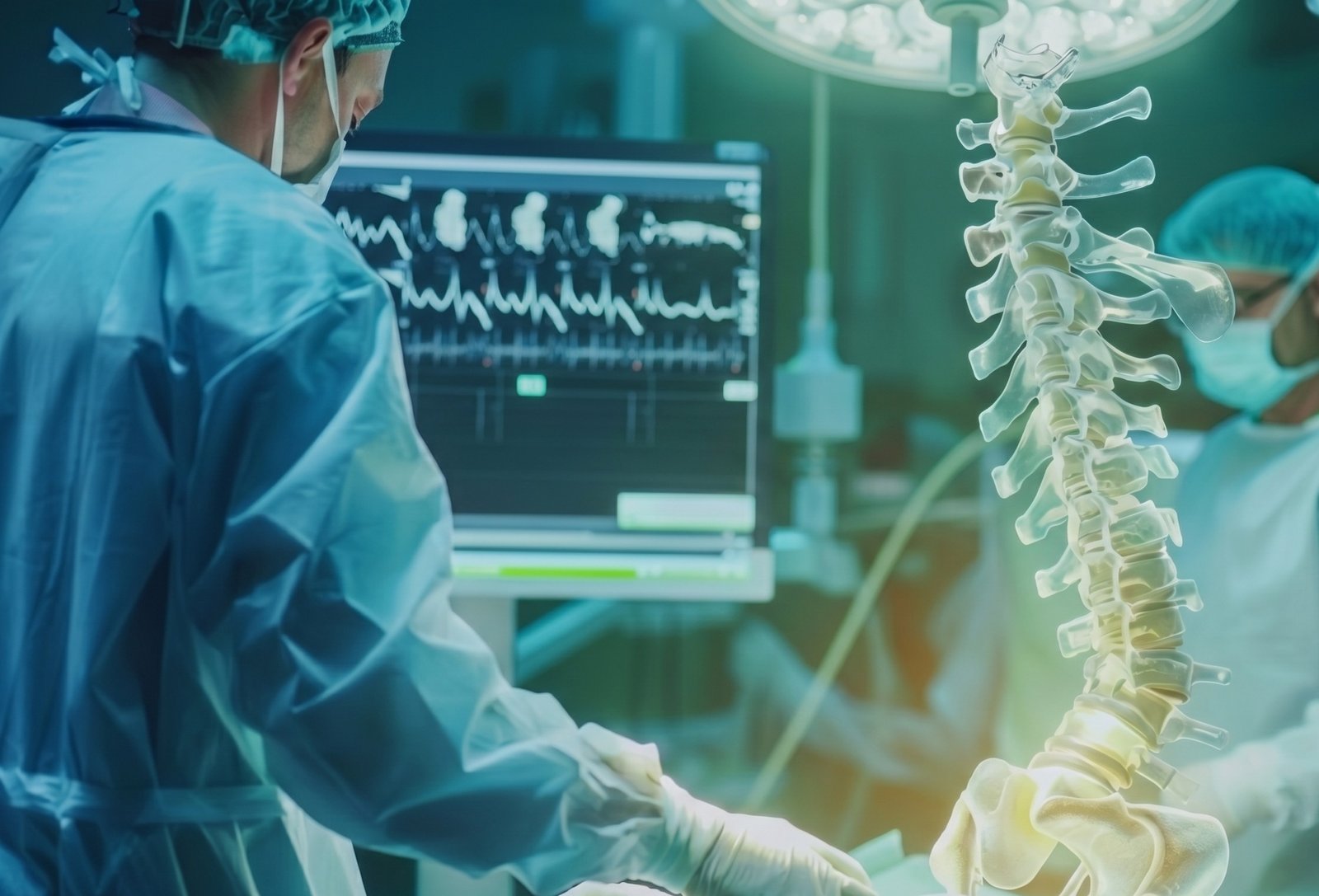
Under high-definition 4K camera magnification and real-time C-Arm fluoroscopic guidance, Dr. Shrutika Bhagat navigates directly to the herniated disc fragment or compressing bone spur, delicately removing only the pathologic tissue while leaving healthy bone, ligaments, and stabilizing spinal architecture 100% intact.
Who is a Candidate for Endoscopic Decompression?
Endoscopic decompression is indicated for patients suffering from spinal nerve compression who haven't found adequate relief from medication or physical therapy.
Lumbar Disc Herniation / Sciatica
L3-L4, L4-L5, or L5-S1 disc protrusions causing shooting pain, tingling, numbness, and burning sensation down the buttocks, thigh, calf, and foot.
Lumbar Canal & Foraminal Stenosis
Narrowing of the spinal nerve exit tunnels causing neurogenic claudication (inability to walk continuous distances without sitting or leaning forward).
Cervical Disc Bulge & Radiculopathy
Neck disc herniation causing severe shooting electrical pain, weakness, and numbness radiating down the shoulder, arm, forearm, and fingers.
Recurrent Disc Prolapse
Re-herniation of disc fragments in patients who had prior open spine surgery, safely accessed through transforaminal keyhole routes without cutting previous scar tissue.
Failed Back Surgery Syndrome (FBSS)
Direct endoscopic camera adhesiolysis and targeted decompression for patients struggling with persistent post-surgical epidural scar fibrosis.
Elderly Patients High-Risk for General Anesthesia
Patients with cardiac disease, diabetes, hypertension, or advanced age who cannot safely undergo general anesthesia or open surgical stress.
Endoscopic Decompression vs. Traditional Open Surgery
| Clinical Parameter | Endoscopic Decompression (Keyhole) | Traditional Open Spine Surgery |
|---|---|---|
| Incision Size | 7 - 8 mm (Stitchless) | 50 - 100 mm (Large Scar) |
| Anesthesia Type | Local Anesthesia + Conscious Sedation | General Anesthesia Required |
| Muscle & Bone Damage | Zero (Muscle Dilation Only) | Extensive Muscle Cutting & Bone Removal |
| Hospital Stay | Same-Day Discharge (Daycare) | 3 to 7 Days Hospitalization |
| Blood Loss | < 5 ml (Negligible) | 100 - 300 ml (May need transfusion) |
| Hardware / Screw Implants | None (Preserves Native Mobility) | Often Requires Screws, Rods & Fusion |
| Post-Op Recovery & Return to Work | 3 to 7 Days | 6 to 12 Weeks of Bed Rest |
How Endoscopic Decompression is Performed
MRI Trajectory Planning
Precise millimeter mapping of the herniated fragment location relative to the nerve root, identifying the optimal transforaminal or interlaminar approach.
Local Anesthesia & Cannulation
Gentle local numbing agent is placed. Under live C-Arm X-ray, a thin guiding wire is advanced safely to the disc space while you remain awake and pain-free.
7mm Working Channel Placement
A pencil-thin 7-8mm cannula gently dilates muscle fibers without cutting any muscle or tissue, providing a direct optical pathway to the spine.
4K High-Definition Visualization
The surgical endoscope is inserted. Continuous sterile saline wash magnifies nerves, blood vessels, and disc fragments on 4K medical monitors.
Targeted Fragmentectomy
Micro-forceps and radiofrequency probes remove only the compressing disc fragment. The freed nerve root is visually confirmed to have full pulsatile mobility.
Stitchless Band-Aid Closure
The cannula is gently withdrawn. No stitches or staples are required—a simple waterproof Band-Aid dressing is applied and you walk in 2 hours.
Endoscopic Decompression Modalities Offered in Patna
Transforaminal Endoscopic Discectomy
Enters through the natural neuroforaminal window from the flank, ideal for L3-L4 and L4-L5 herniations with zero bone removal.
Interlaminar Endoscopic Discectomy
Enters through the interlaminar space from the posterior back, ideal for L5-S1 disc prolapse protected by high iliac crest bones.
Unilateral Biportal Endoscopy
Uses two tiny portals (one for camera, one for instruments) providing superior freedom for severe central spinal stenosis decompression.
Posterior Cervical Discectomy
Keyhole access from the back of the neck for herniated cervical discs, avoiding anterior neck surgery and bone fusion plates.
Common Questions on Endoscopic Decompression
Why Choose Dr. Shrutika for Endoscopic Decompression in Patna
- Targeted Fragmentectomy: Only the herniated disc fragment compressing the sciatic nerve is removed, keeping the healthy cushioning disc structure completely intact.
- Zero Spinal Fusion Required: Avoids metallic implants, cages, or spinal hardware, preventing long-term adjacent segment disc degeneration and stiffness.
- Minimal Blood Loss & Zero Muscular Trauma: The transforaminal micro-port dilates muscles gently without cutting, completely preventing post-operative scar tissue fibrosis.
- Immediate Sciatica Relief: Direct unpinching of the trapped spinal root provides instant relief from shooting leg pain, calf tingling, and foot numbness.
- Rapid Walking in 2 Hours: Conducted under local anesthesia in daycare; patients stand, walk, and are discharged home comfortably on the same day.
- Proven 95%+ Success Rate: Consistently high clinical success and positive patient outcomes for L4-L5 and L5-S1 lumbar disc herniations.


Don't Let Chronic Pain Control Your Life.
Take the first step toward understanding your pain and exploring modern, evidence-based minimally invasive relief with Dr. Shrutika Bhagat.