Lumbar Spondylosis Treatment in Patna
Relief from chronic lower back arthritis, morning stiffness, bone spurs & walking leg fatigue through precision facet radiofrequency ablation & non-surgical spine care.
Understanding Lumbar Spondylosis (Lower Back Osteoarthritis)
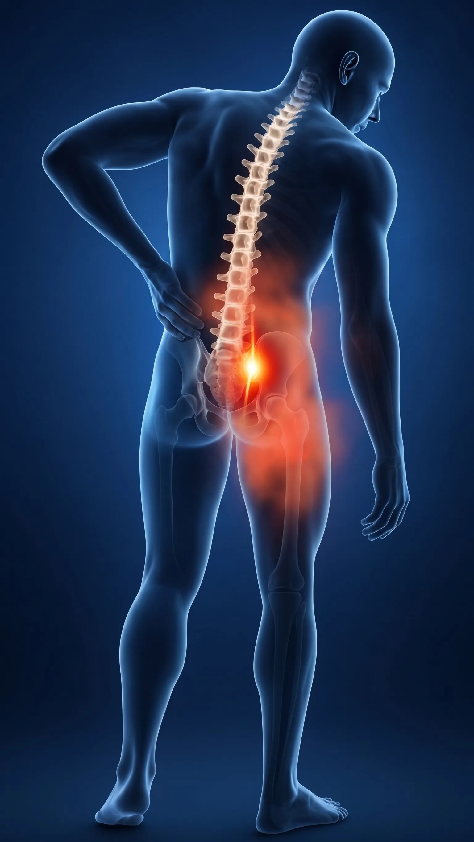
Lumbar Spondylosis is an umbrella clinical term describing age-related and mechanical wear-and-tear degeneration of the lower lumbar spine (L1 to S1 vertebrae). It involves progressive changes in intervertebral discs, facet joints, ligamentum flavum, and bony vertebral margins.
The lower back bears the majority of upper body weight while facilitating bending and twisting. As intervertebral discs dehydrate and lose height over time, biomechanical load shifts onto the posterior facet joints. This causes cartilage breakdown, joint friction, and the growth of compensatory Osteophytes (Bone Spurs).
This degenerative cascade typically presents in three clinical forms:
- Axial Facet Joint Pain: Deep lower back aching and morning stiffness that worsens with standing or bending backward.
- Lumbar Radiculopathy (Sciatica): Osteophytes or bulging disc margins compressing exiting nerve roots, causing shooting leg pain.
- Spinal Canal Stenosis: Thickened ligaments and bone spurs narrowing the central canal, causing cramping and heaviness in both legs after walking (Neurogenic Claudication).
Interventional Spine Care targets the exact arthritic joint or compressed nerve root with sub-millimeter precision under live C-Arm fluoroscopy, eliminating pain without major open spinal fusion surgeries or metal implants.
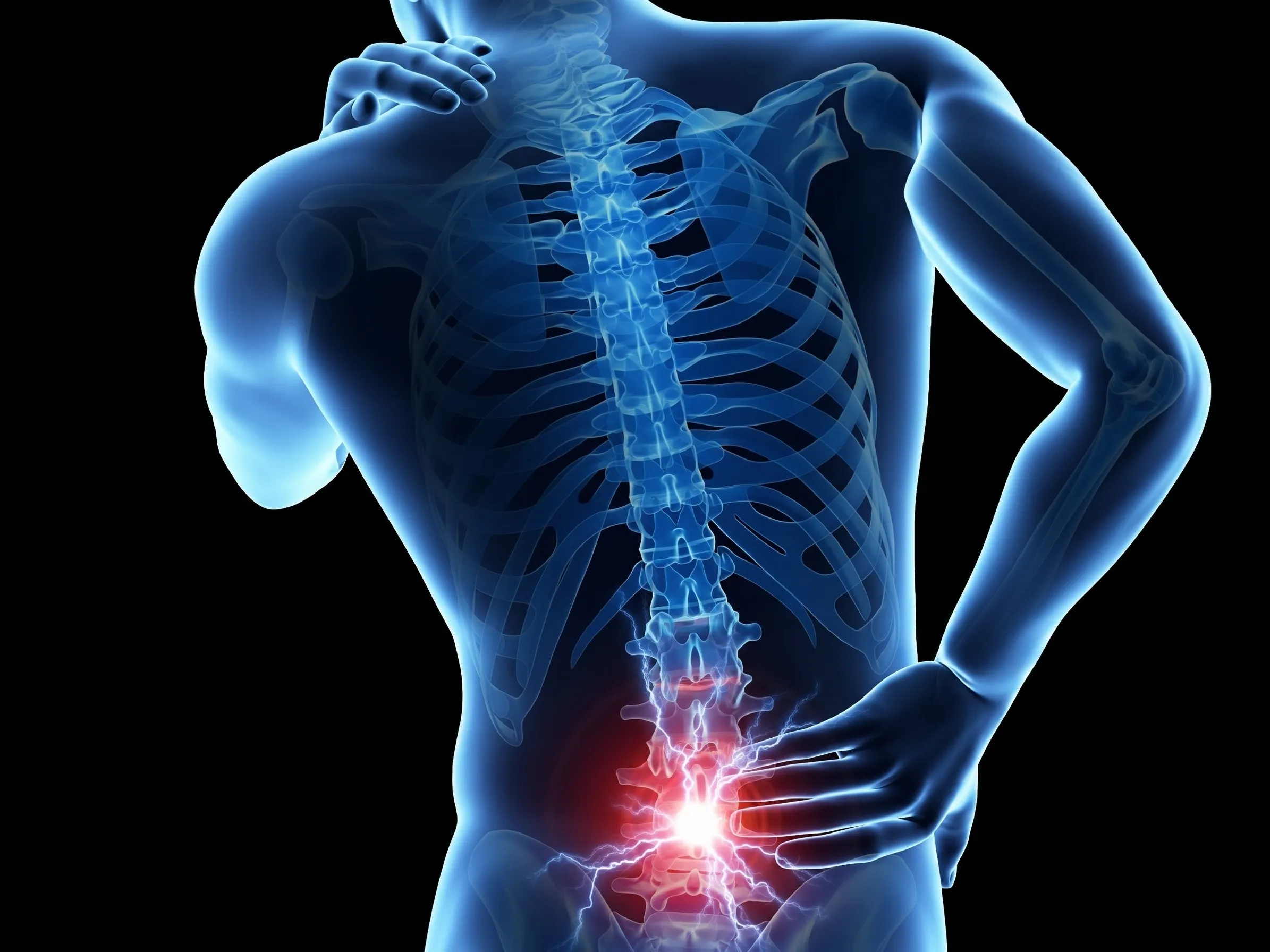
How Lumbar Spondylosis Manifests
Symptoms vary depending on whether the primary degeneration involves facet joints, exiting nerve roots, or the central spinal canal.
1. Axial Facet Joint Arthritis
Deep, aching lower back pain and stiffness upon waking (lasting <30 minutes). Aggravated by standing upright, walking, or bending backward (extension), and relieved by sitting down or bending forward.
- Primary Cause: Cartilage loss in L4-L5 & L5-S1 facet joints.
- Referred Pain: Buttocks, groin, and upper posterior thigh.
2. Spondylotic Radiculopathy (Sciatica)
Sharp shooting pain, burning, tingling, and pins-and-needles traveling down the buttock, thigh, calf, or foot caused by arthritic bone spurs narrowing the neural exit foramen and pinching nerve roots.
- Primary Cause: Foraminal stenosis compressing L4, L5, or S1 nerves.
- Associated: Foot drop, weak big toe, or lost ankle reflex.
3. Spinal Stenosis & Claudication
Heaviness, cramping, and fatigue in both legs after walking a fixed distance (100–300 meters), forcing the patient to sit down or lean forward (the classic "Shopping Cart Sign").
- Primary Cause: Thickened ligamentum flavum & central canal narrowing.
- Relieved by: Sitting or bending the spine forward.
Lumbar Spondylosis Levels & Symptoms
The exact location of your lower back pain and leg symptoms corresponds directly to the degenerated lumbar segment.
| Lumbar Segment | Facet & Nerve Level | Pain & Stiffness Distribution | Functional & Reflex Impact |
|---|---|---|---|
| L1 – L3 Levels | L1, L2, L3 Nerves | Upper lumbar backache, groin pain, and anterior upper thigh aching | Stiffness when getting out of bed or turning in sleep; Hip flexor weakness |
| L3 – L4 Level | L4 Nerve Root | Mid-lower back, hip, front of the thigh, knee, and inner lower leg | Weak knee straightening (quadriceps); Diminished Patellar Knee Jerk reflex |
| L4 – L5 Level | L5 Nerve Root | Lower back, buttock, outer thigh, outer calf, top of foot, and big toe | Difficulty walking on heels (Foot Drop risk); Weak big toe extension |
| L5 – S1 Level | S1 Nerve Root | Lumbosacral junction, buttock, back of thigh, calf, heel, and little toe | Difficulty standing on tiptoes; Diminished Achilles Ankle Jerk reflex |
Our Diagnostic Protocol for Lumbar Spondylosis
Combining targeted physical examination with cutting-edge radiology guarantees precise identification of the arthritic pain generator.
Targeted Physical Exam
Kemp's extension test for facet joint arthropathy, Straight Leg Raise (SLR), SI joint loading tests, and walking claudication timing.
Dynamic Spine X-Rays
Standing flexion and extension views to reveal disc space narrowing, osteophyte bridges, and subtle spondylolisthesis slipping.
3-Tesla MRI Spine
Gold-standard imaging detailing facet joint arthrosis, ligamentum flavum thickening, canal stenosis caliber, and nerve crowding.
Diagnostic Facet Block
Targeted micro-injection of local anesthetic over the lumbar medial branch nerves under C-Arm to confirm joint pain before RFA.
Non-Surgical Interventions for Lumbar Spondylosis
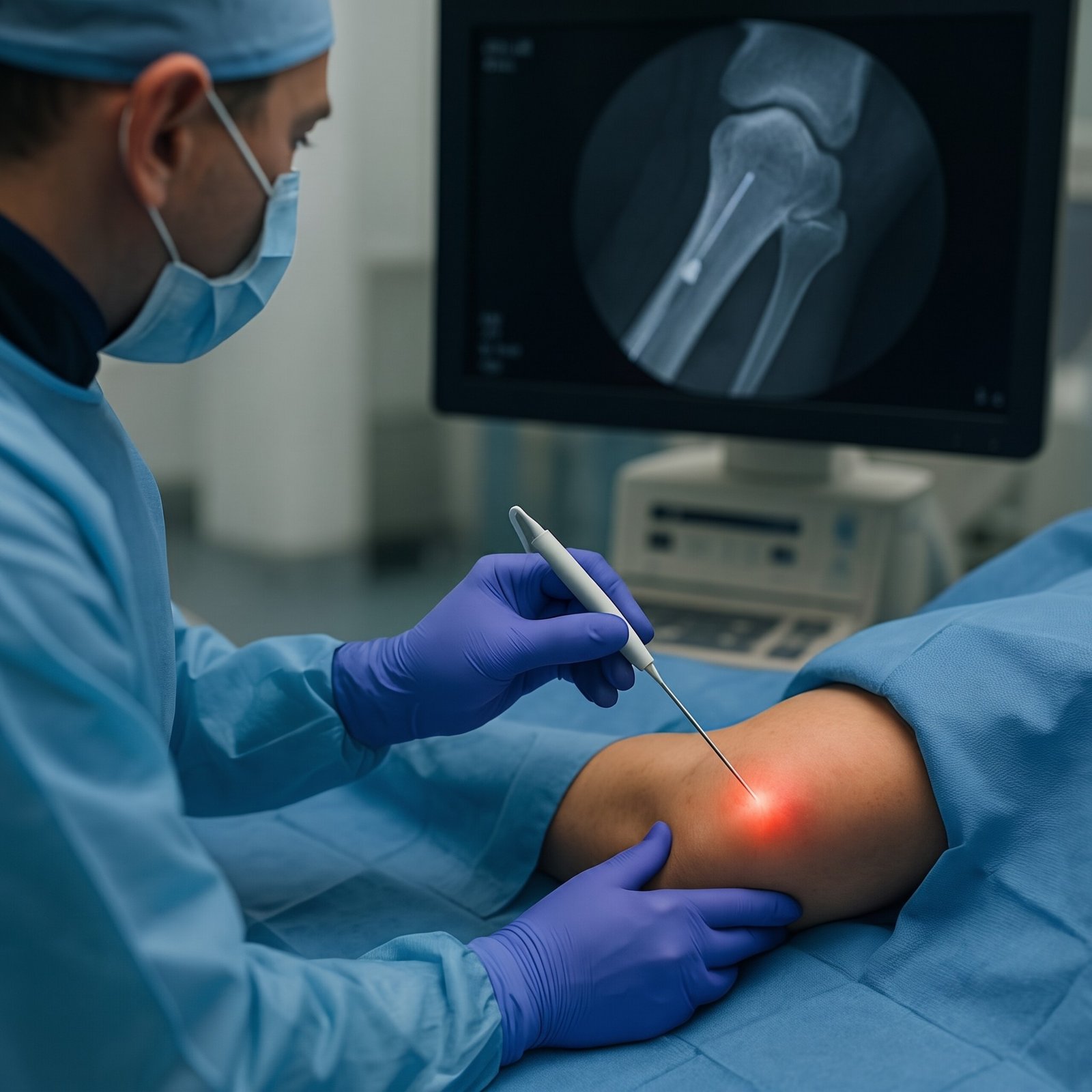
Daycare interventional procedures performed under live fluoroscopic C-Arm and ultrasound guidance by Dr. Shrutika Bhagat.
 Long-Term Relief
Lumbar Facet Radiofrequency (RFA)
C-Arm fluoroscopy-guided thermal denervation applied to the medial branch sensory nerves supplying arthritic facet joints, disabling pain signals and providing 12 to 24 months of sustained lower back relief.
- Deactivates chronic lower back arthritic pain
- 100% motion preservation, zero spinal fusion
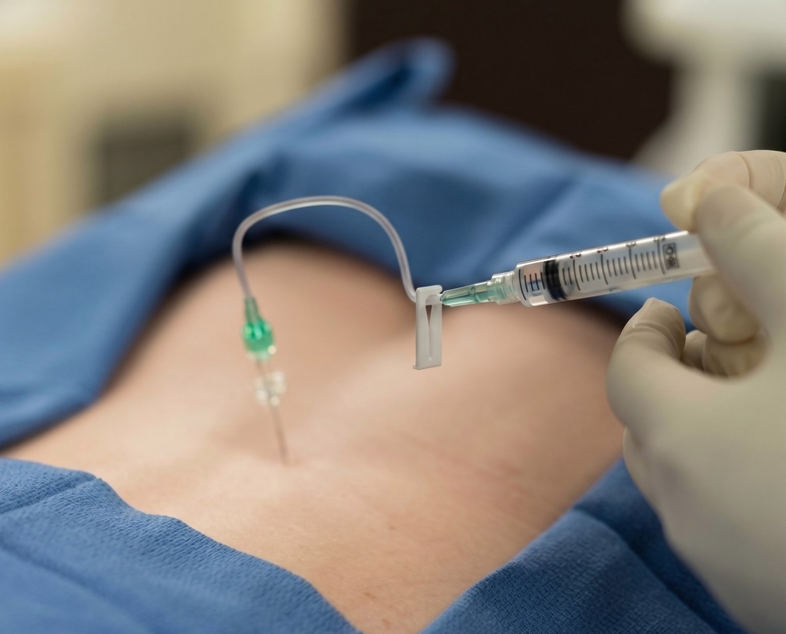 Image-Guided
Lumbar Epidural Injection (TFESI)
Precision fluoroscopy-guided injection delivering concentrated anti-inflammatory medication directly into the epidural space adjacent to the compressed nerve root, rapidly clearing radiating sciatica and leg pain.
- Rapid relief from shooting leg pain and numbness
- Calms nerves choked by bone spurs & stenosis
 Pelvic-Spine Balance
SI Joint Injection & RFA
Lumbar spondylosis frequently shifts mechanical load down into the sacroiliac (SI) joints. Image-guided injections and lateral branch radiofrequency denervation eliminate deep buttock and pelvic aching.
- Solves deep buttock & sitting agony
- Restores harmonious lumbopelvic dynamics
 Targeted Adhesiolysis
Epidural Catheter Neuroplasty
A specialized steerable micro-catheter gently navigates into the lumbar epidural canal under C-Arm imaging to break down fibrotic scar bands and chronic adhesions entrapping nerve roots in spondylosis.
- Mechanical release of bound spinal nerves
- Highly effective for chronic spondylotic pain
 Biologic Healing
Spine PRP & Facet Biotherapy
Autologous concentrated platelet growth factors injected under live imaging into worn lumbar facet capsules and supportive spinal ligaments to stimulate cellular cartilage repair and reinforce spinal stability.
- 100% natural, patient's own healing biology
- Tightens loose, arthritic lumbar motion segments
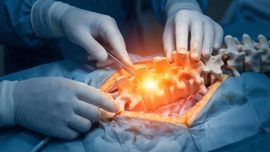 Keyhole Procedure
Keyhole Endoscopic Decompression
Ultra-minimally invasive stitchless decompression (TELD / IELD) through a tiny 7–8mm port under local anesthesia. An HD camera visualizes the narrowed canal, trimming bone spurs without metal spinal fusion.
- Zero metal rods, screws, or spinal fusion
- Walk home within 2 hours of procedure
Open Spine Fusion (TLIF) vs. Interventional Care
Why non-surgical interventional procedures and Radiofrequency Ablation are the gold standard for lumbar spondylosis.
| Clinical Feature | Open Spinal Fusion (TLIF / PLIF) | Interventional Spine Care & RFA |
|---|---|---|
| Anesthesia Required | General Anesthesia (higher medical risks) | Local Anesthesia / Mild Conscious Sedation |
| Incision & Stitches | Large 3 – 5 inch incision with extensive stitches | Micro-needle punctures (Stitchless daycare) |
| Natural Spine Mobility | Permanently fuses spine (Loss of natural bending) | 100% preserves natural flexibility & lumbar movement |
| Metal Implants & Screws | 4 to 8 titanium pedicle screws, rods & cages | Zero metal implants or artificial hardware inserted |
| Adjacent Segment Disease Risk | High risk (Excess stress shifts to adjoining discs) | Zero adjacent segment overload risk |
| Hospital Stay & Work Return | 4 to 7 days hospital stay; 2-3 months bed rest | Daycare: Discharged in 2 hours; work in 2–4 days |

Dr. Shrutika Bhagat
MBBS, MD, FIRA, FIAPM | Interventional Pain & Spine Endoscopy
Dr. Shrutika Bhagat brings super-specialized training in full-endoscopic spine surgery and image-guided interventional pain management, having successfully treated thousands of patients with severe back pain, disc prolapse, sciatica, nerve compression, and joint arthritis in Patna.
Lumbar Spondylosis FAQs
Clear clinical answers regarding lower back arthritis, facet radiofrequency ablation, spinal stenosis, and recovery.
Don't Let Chronic Pain Control Your Life.
Take the first step toward understanding your pain and exploring modern, evidence-based minimally invasive relief with Dr. Shrutika Bhagat.