SI Joint Pain Treatment in Patna
Relief from deep buttock pain, sitting discomfort & pelvic instability through precision C-Arm guided SI joint injections, cooled lateral branch radiofrequency & PRP biotherapy.
Understanding Sacroiliac (SI) Joint Dysfunction
The Sacroiliac (SI) Joint connects the triangular base of your spine (the sacrum) to the pelvic bones (ilium). It acts as a massive shock absorber, transferring rotational and vertical forces between the upper body and the legs.
SI joint dysfunction is responsible for up to 15% to 30% of all lower back and buttock pain cases, and is frequently misdiagnosed as a lumbar disc herniation or sciatica. It can be triggered by uneven gait, pelvic ligament laxity during pregnancy, prior lumbar fusion surgery (which shifts stress onto the SI joint), or inflammatory spondyloarthropathies (Ankylosing Spondylitis).
The posterior sacroiliac joint and its dense stabilizing ligaments are innervated by the Sacral Lateral Branch Nerves (S1, S2, S3).
Cooled Radiofrequency Ablation (RFA) and regenerative PRP biotherapy provide lasting, targeted relief by deactivating pain-transmitting lateral branches and biologically tightening lax pelvic ligaments without open joint fusion.
Symptoms of Sacroiliac Joint Pain
Identifying the characteristic pain triggers of SI joint dysfunction to differentiate it from lumbar disc herniation.
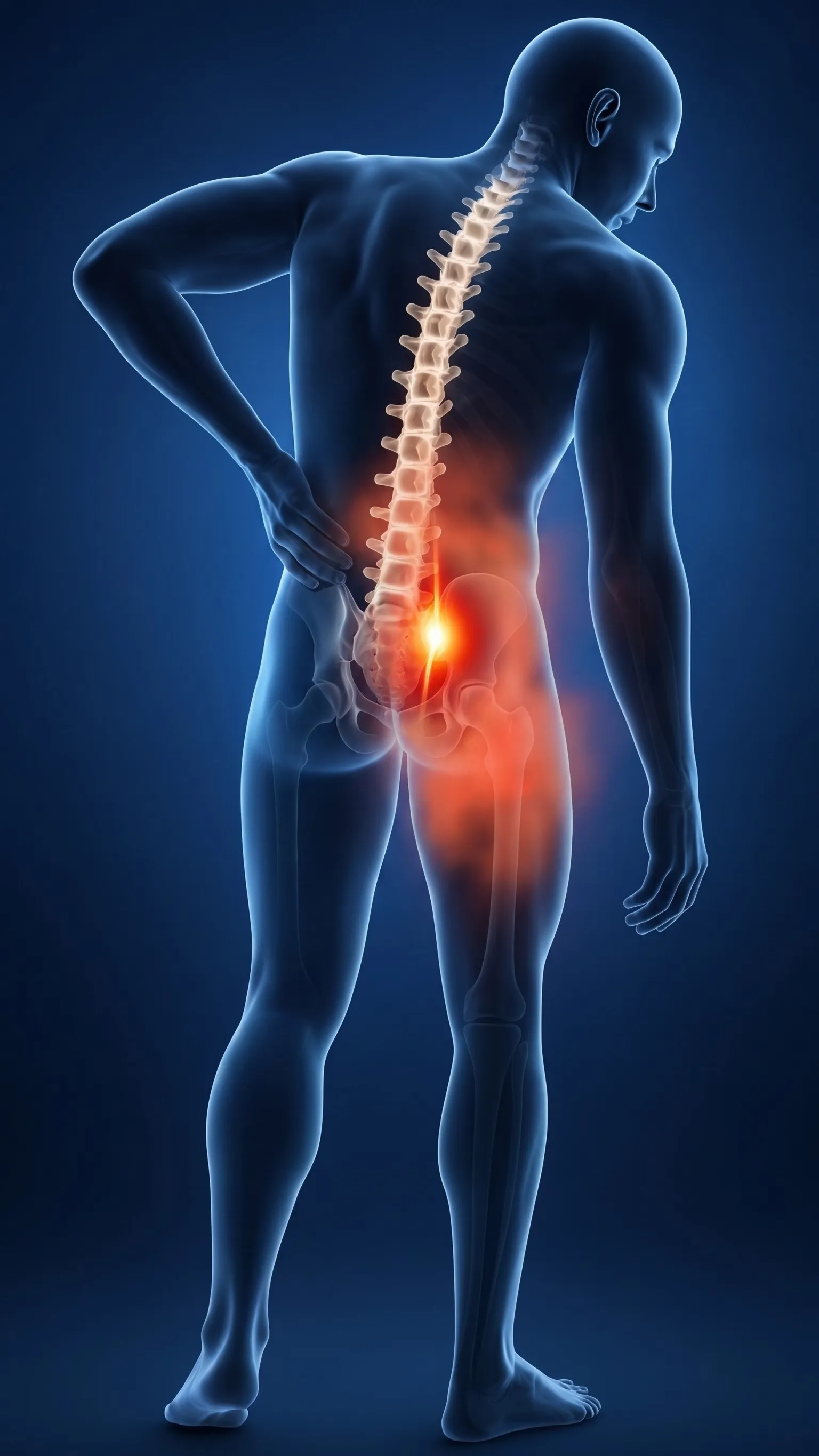
Pain on Sitting & Shifting Weight
Deep aching on one side of the buttock, worsening when sitting in a car, sitting cross-legged, or shifting body weight onto one side.
Pain on Rising & Climbing Stairs
A sharp catch in the pelvic dimple when transitioning from sitting to standing, climbing stairs, or rolling over in bed at night.
Groin & Posterior Thigh Referral
Referred pain radiating into the posterior thigh and groin that mimics sciatica, but stops above the knee and lacks true neurological numbness.
Our 4-Step Diagnostic Protocol for SI Joint Pain
Using validated clinical provocative maneuvers and precision fluoroscopic testing to establish the exact diagnosis.
Provocative Tests
Battery of 5 physical provocative tests (FABER/Patrick's, Gaenslen's, Distraction, Compression, Thigh Thrust). 3+ positive confirms SI origin.
Digital Pelvis X-Rays
Standing Ferguson view pelvic X-rays to detect joint space narrowing, subchondral sclerosis, or pelvic obliquity.
3T MRI SI Joints
STIR sequences to detect active bone marrow edema in Sacroiliitis (Ankylosing Spondylitis) vs mechanical degenerative wear.
Diagnostic Block
Gold Standard: Fluoroscopy-guided intra-articular test injection. >75% relief confirms SI joint pathology.
Non-Surgical Interventions for SI Joint Pain
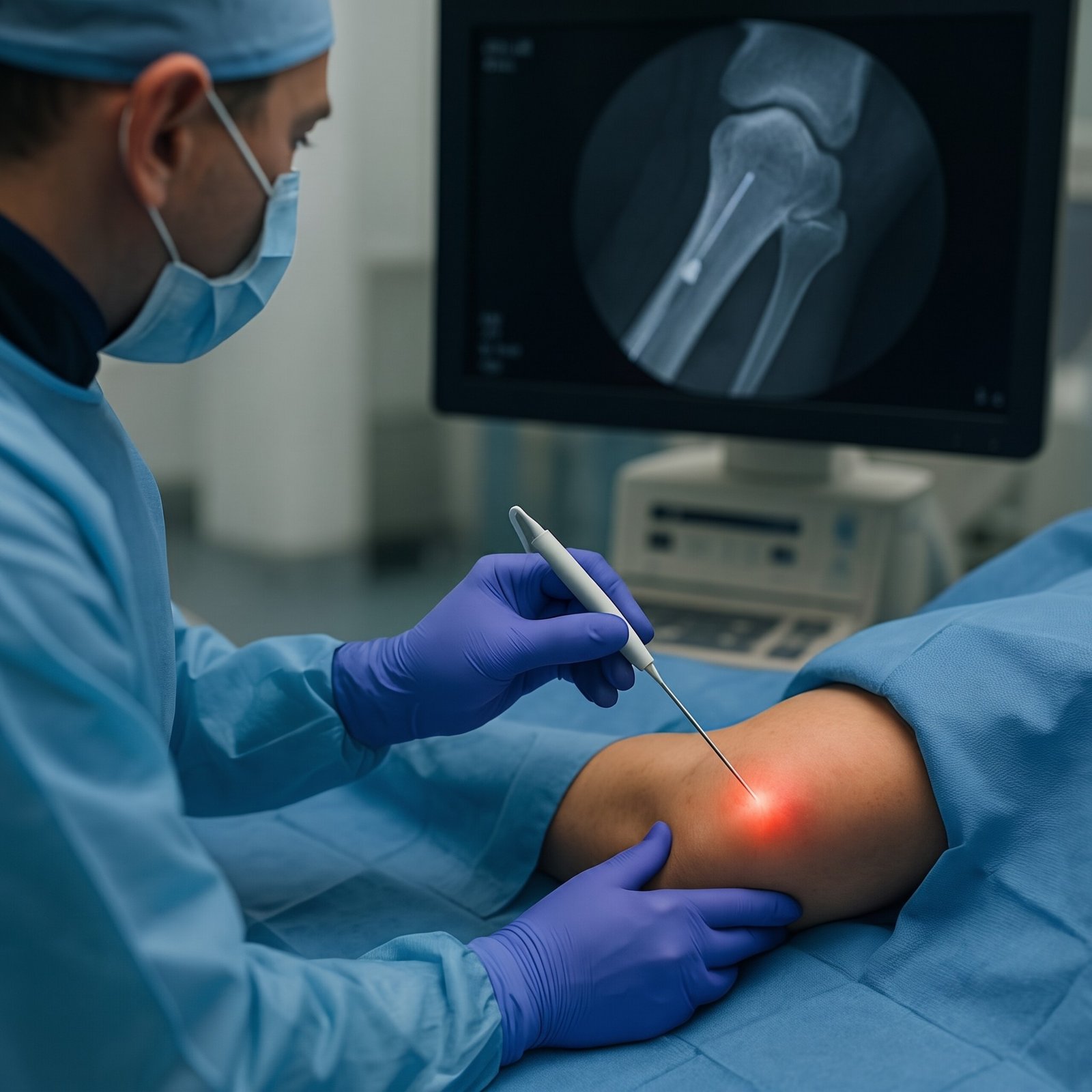
Daycare needle procedures performed under live fluoroscopic C-Arm guidance by Dr. Shrutika Bhagat in Patna.
Image-Guided
Intra-Articular SI Joint Injections
C-Arm fluoroscopy-guided precision injection into the narrow SI joint space to extinguish severe synovial inflammation and restore sitting tolerance.
- Fast relief from acute sacroiliac inflammation
- Daycare procedure under local anesthesia
 12-24 Mos Relief
Sacral Lateral Branch RFA
Cooled radiofrequency thermal neurolysis deactivating the sensory lateral branch nerves (S1–S3) and L5 dorsal ramus, shutting down chronic buttock and pelvic pain for 1 to 2 years.
- Long-lasting relief without open joint fusion
- Preserves all leg motor strength & walking
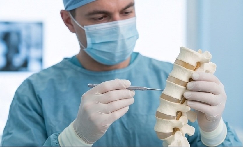 Biologic Repair
Autologous SI Ligament PRP
Platelet-rich plasma growth factors injected under ultrasound/fluoroscopy directly into lax dorsal sacroiliac and iliolumbar ligaments to biologically tighten and stabilize hypermobile joints.
- 100% natural healing growth factors
- Ideal for post-pregnancy & ligament laxity
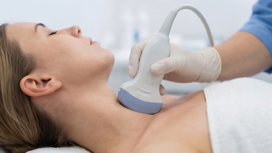 Nerve Decompression
Piriformis & Sciatic Hydrodissection
High-resolution ultrasound-guided release of tense piriformis muscle spasms and scarred fascial bands compressing the sciatic nerve adjacent to the SI joint.
- Stops shooting buttock and thigh pain
- Restores painless sitting in chairs
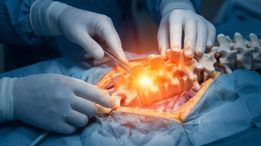 Keyhole Surgery
Endoscopic SI Joint Denervation
Stitchless keyhole endoscopy through a 7mm portal under direct HD camera vision to ablate chronic sensory lateral branch nerves when conventional measures fail.
- Direct visual visualization of nerve fibers
- Zero bone destruction or metal screws
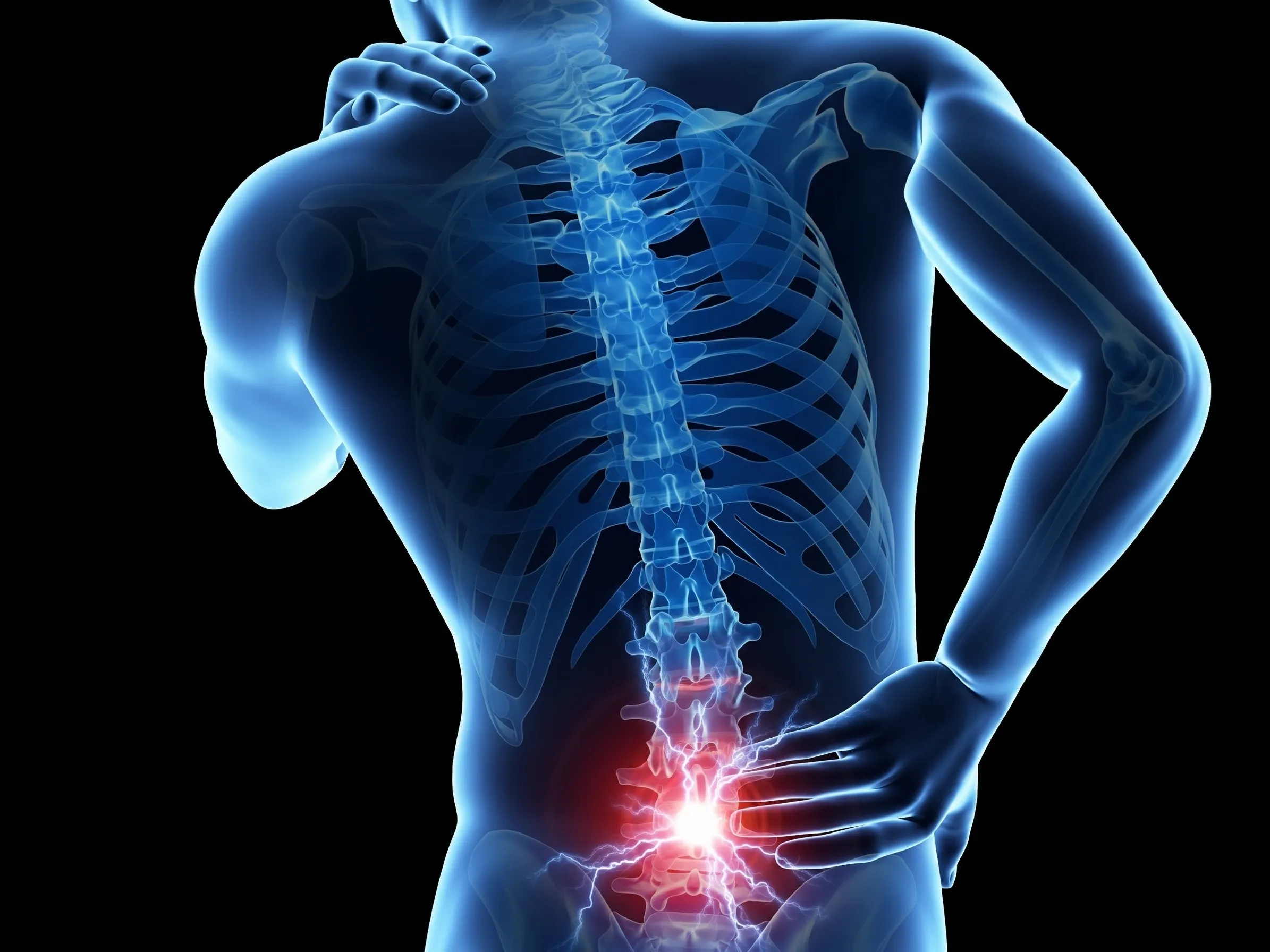 Rehabilitation
Pelvic Alignment & Core Rehab
Customized pelvic belt stabilization, leg length discrepancy correction, and deep core strengthening (transversus abdominis and gluteal activation).
- Restores symmetrical walking biomechanics
- Prevents recurrent pelvic torsion
Open SI Joint Fusion Surgery vs. Daycare Cooled RFA
Why non-surgical interventional denervation is the preferred standard for chronic sacroiliac pain.
| Clinical Feature | Open SI Joint Fusion Surgery | Daycare Cooled Lateral Branch RFA |
|---|---|---|
| Anesthesia Type | General Anesthesia | Local Anesthesia only (Conscious & safe) |
| Incision & Screws | Large open cut with 3 large titanium fusion screws | Pinhole needle entry (Zero metal implants) |
| Pelvic Joint Mobility | Permanently fuses and eliminates joint shock-absorption | Preserves 100% natural pelvic biomechanics |
| Weight-Bearing Restrictions | 6 to 12 weeks of non-weight-bearing crutches | Immediate full weight-bearing on the same day |
| Hospital Stay | 3 to 5 days hospital admission | Daycare: Discharged in 2 to 3 hours |

Dr. Shrutika Bhagat
MBBS, MD, FIRA, FIAPM | Interventional Pain & Spine Endoscopy
Dr. Shrutika Bhagat brings super-specialized training in full-endoscopic spine surgery and image-guided interventional pain management, having successfully treated thousands of patients with severe back pain, disc prolapse, sciatica, nerve compression, and joint arthritis in Patna.
Sacroiliac (SI) Joint FAQs
Clinical answers regarding SI joint dysfunction, sitting pain, cooled radiofrequency, and PRP prolotherapy.

Don't Let Chronic Pain Control Your Life.
Take the first step toward understanding your pain and exploring modern, evidence-based minimally invasive relief with Dr. Shrutika Bhagat.