Slip Disc Treatment in Patna
Relief from herniated disc pain, sciatica, and nerve compression through precision image-guided nerve blocks, ozone discolysis & stitchless keyhole spine endoscopy.
Understanding Slip Disc & Herniated Disc Prolapse (PIVD)
An intervertebral disc acts as a shock-absorbing cushion between each bony vertebra of your spine. Each disc consists of two main parts: a tough, fibrous outer ring (Annulus Fibrosus) and a soft, jelly-like inner core (Nucleus Pulposus).
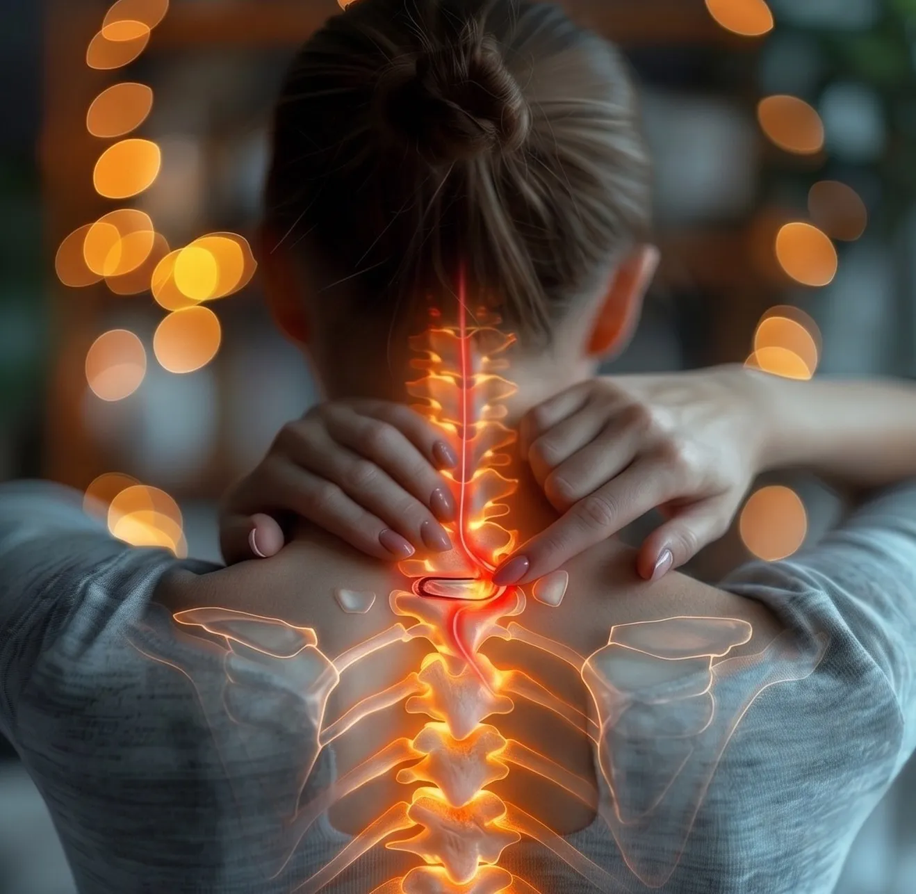
A Slip Disc (medically termed Herniated Disc or Prolapsed Intervertebral Disc - PIVD) occurs when the outer ring weakens, tears, or fissures, allowing the inner gel to bulge or extrude outwards into the spinal canal or exit neural foramen.
This herniated disc material causes severe agony through two distinct mechanisms:
- Mechanical Compression: Physical crowding and pinching of traversing spinal nerve roots (triggering sciatica, shooting leg pain, or arm weakness).
- Chemical Radiculitis: The leaking nucleus gel secretes acidic inflammatory enzymes (Phospholipase A2, TNF-α) that severely inflame and swell the surrounding nerves.
Advanced Interventional Spine Care neutralizes both the chemical inflammation and mechanical compression under live C-Arm guidance—enabling full recovery without major open surgery, cutting bone, or inserting metal screws.
The 4 Stages of Slip Disc Progression
Understanding your MRI grade allows Dr. Shrutika Bhagat to customize the most effective minimally invasive intervention.
Disc Bulge / Degeneration
The disc dehydrates and weakens with age or strain. The outer ring bulges uniformly outward (like a flattened tire) without tearing.
- Symptoms: Dull lower back ache, stiffness upon waking.
Disc Protrusion
The nucleus gel pushes focal-wise into partial tears within the inner annular layers, forming a localized out-pocketing that contacts the thecal sac.
- Symptoms: Sharp back pain radiating to buttock or upper thigh.
Disc Extrusion
The tough outer ring ruptures completely. The gel-like nucleus escapes into the epidural space but remains connected to the main disc body.
- Symptoms: Severe sciatica, electric shocks down calf, foot numbness.
Disc Sequestration
Extruded disc material breaks away completely, forming a "free fragment" that migrates up or down inside the spinal canal, severely choking nerve roots.
- Symptoms: Intractable leg agony, motor weakness (foot drop), muscle wasting.
Where Does Slip Disc Occur?
Over 95% of slip discs occur in the lower back (Lumbar) or the neck (Cervical), producing distinct symptom pathways.
Lumbar Slip Disc (Lower Back)
The lumbar spine bears the greatest biomechanical load. Herniations predominantly happen at L4-L5 and L5-S1, irritating the sciatic nerve.
Pain & numbness down outer thigh, outer shin, top of the foot, and big toe. Risk of Foot Drop (inability to lift foot while walking).
Pain radiating down back of thigh, calf, heel, and outer sole of the foot. Weakness when standing on tiptoes and lost ankle jerk reflex.
Pain in front of thigh and knee, with quadriceps weakness (difficulty climbing stairs or straightening the knee).
Cervical Slip Disc (Neck)
The flexible neck column is vulnerable to mobile screen strain ("Text Neck") and posture stress. Herniations commonly strike at C5-C6 and C6-C7.
Severe neck stiffness, pain shooting down shoulder, bicep, outer forearm, thumb, and index finger. Biceps muscle weakness.
Pain traveling down back of the arm, triceps, into the middle finger. Triceps weakness and impaired grip strength.
Numbness and pins-and-needles in the little finger and ring finger, accompanied by hand clumsiness and dropping objects.
Our Diagnostic Protocol for Slip Disc
Precise diagnosis ensures that only the exact pain-generating disc level is treated without unnecessary invasive interventions.
Neurological Testing
Straight Leg Raise (SLR test), Lasegue sign, Spurling test (for neck), sensory dermatome mapping, and muscle power grading.
Dynamic Spine X-Rays
Weight-bearing flexion & extension views to evaluate disc space collapse, vertebral alignment, and segmental instability.
3-Tesla MRI Spine
High-definition MRI reveals exact disc herniation dimensions, annular fissures, canal stenosis, and exiting nerve root impingement.
Diagnostic Test Block
Targeted micro-injection of local anesthetic under live C-Arm fluoroscopy to definitively verify the offending disc before procedure.
Non-Surgical & Keyhole Treatments for Slip Disc
Daycare interventional procedures performed under real-time C-Arm fluoroscopic imaging by Dr. Shrutika Bhagat in Patna.
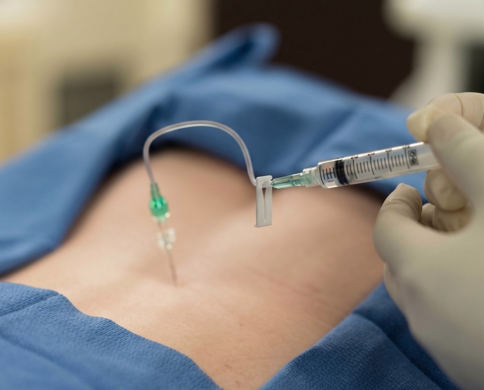 Image-Guided
Transforaminal Epidural (TFESI)
C-Arm fluoroscopy-guided precision injection targeting the exit foramen directly adjacent to the compressed nerve root, delivering high-potency anti-inflammatory medication to rapidly alleviate sciatica and back pain.
- Fast relief from acute shooting leg & arm pain
- Accelerates natural disc resorption
 Keyhole Procedure
Keyhole Endoscopic Discectomy
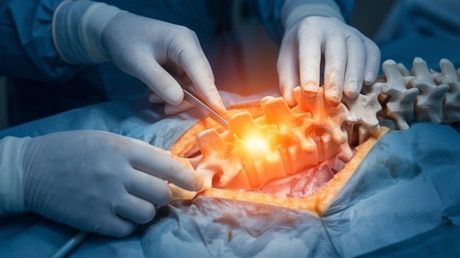
Ultra-minimally invasive stitchless decompression (TELD / IELD) through a 7–8mm port under local anesthesia. An HD camera visualizes the pinched nerve and precisely extracts only the extruded disc fragment.
- Zero bone cutting or metal implants
- Walk home comfortably within 2 hours
Non-Surgical Shrinkage
Ozone Nucleolysis (Discolysis)
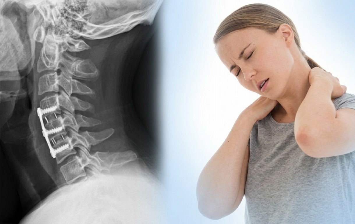
A fine needle introduces medical ozone-oxygen gas directly into the nucleus pulposus under fluoroscopic guidance. Ozone oxidizes proteoglycans, causing the herniated disc to shrink and decompress the nerve.
- Daycare needle procedure, 0 incision
- High success rate for contained disc bulges
 Targeted Adhesiolysis
Epidural Catheter Neuroplasty
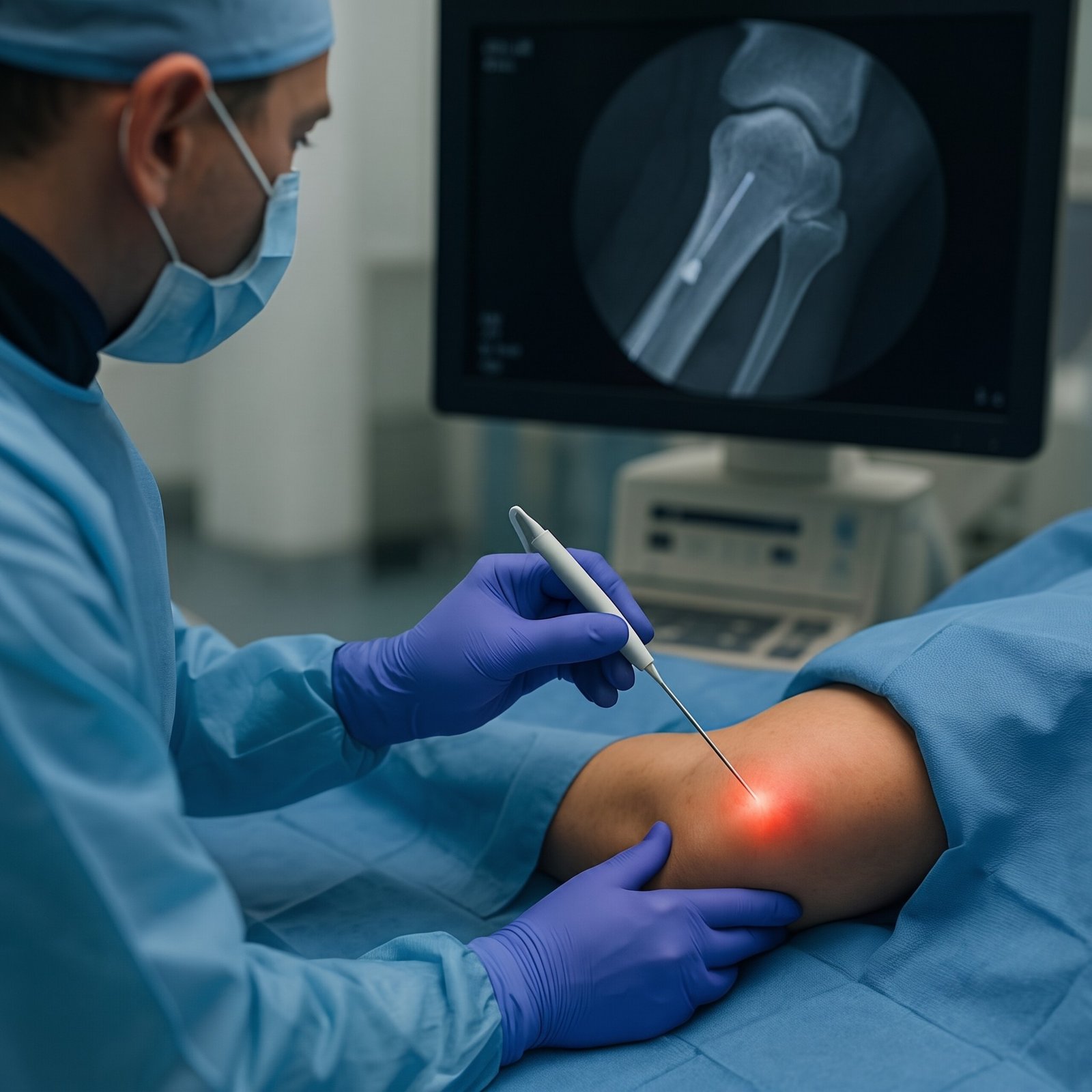
A steerable micro-catheter (Racz catheter) gently breaks down fibrotic adhesions, epidural scar tissue, and inflammatory bands entrapping the nerve root, highly effective for chronic or recurrent slip disc pain.
- Mechanical release of entrapped nerves
- Solves persistent post-operative scarring
 Neuromodulation
Pulsed Radiofrequency (PRF) of DRG
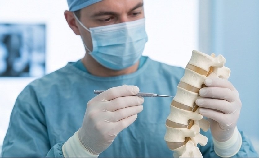
Application of non-destructive pulsed electrical fields to the Dorsal Root Ganglion (DRG), resetting hypersensitive pain transmission pathways and providing 12 to 24 months of sustained relief.
- Neuromodulates chronic nerve hypersensitivity
- 100% safe, zero motor nerve injury
 Biologic Healing
Spine PRP & Annular Biotherapy
Autologous concentrated platelet growth factors injected under precision imaging into the torn outer disc annulus and supportive spinal ligaments to accelerate cellular repair and seal disc tears naturally.
- 100% natural, uses patient's own healing biology
- Strengthens weakened outer disc ring
Open Spine Surgery vs. Interventional Keyhole Care
Why modern interventional spine care has replaced traditional open spine fusion surgeries for slip disc.
| Feature | Traditional Open Spine Surgery | Interventional Keyhole / Endoscopy |
|---|---|---|
| Anesthesia | General Anesthesia (higher medical risk) | Local Anesthesia / Conscious Sedation |
| Incision Size | Large 3 – 5 inch incision with stitches | Tiny 7–8mm keyhole or needle-point (Stitchless) |
| Bone & Muscle Damage | Extensive muscle cutting & bone removal (laminectomy) | Zero muscle cutting, preserves natural spine anatomy |
| Implants / Screws | Often requires metal screws, plates & cages | Zero metal implants or artificial hardware |
| Hospital Stay | 3 to 7 days hospital admission | Daycare: Discharged within 2 to 4 hours |
| Recovery & Work Return | 6 to 12 weeks of restricted mobility | Immediate walking; resume routine in 2–5 days |
| Post-Surgical Scarring Risk | High risk of Failed Back Surgery Syndrome (FBSS) | Minimal scar tissue formation |
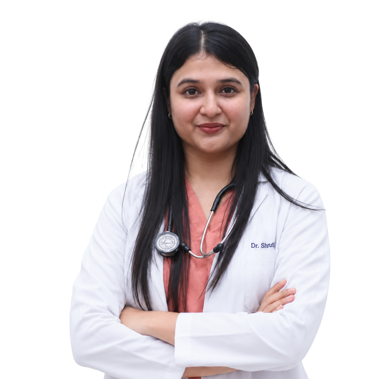
Dr. Shrutika Bhagat
MBBS, MD, FIRA, FIAPM | Interventional Pain & Spine Endoscopy
Dr. Shrutika Bhagat brings super-specialized training in full-endoscopic spine surgery and image-guided interventional pain management, having successfully treated thousands of patients with severe back pain, disc prolapse, sciatica, nerve compression, and joint arthritis in Patna.
Slip Disc FAQs
Clear clinical answers regarding slip disc recovery, non-surgical treatment options, and keyhole procedures.
Don't Let Chronic Pain Control Your Life.
Take the first step toward understanding your pain and exploring modern, evidence-based minimally invasive relief with Dr. Shrutika Bhagat.