Spinal Canal Stenosis Treatment in Patna
Relief from walking leg fatigue, cramping, numbness & spinal canal narrowing through targeted image-guided epidural neuroplasty & stitchless keyhole endoscopy.
Understanding Spinal Canal Stenosis & Neurogenic Claudication
The Spinal Canal is the central bony and ligamentous tunnel traversing the vertebrae, protecting the spinal cord and the bundle of nerve roots traveling to your legs (Cauda Equina).
Spinal Canal Stenosis occurs when this neural passage becomes abnormally narrowed due to age-related wear, hypertrophied (thickened) Ligamentum Flavum, arthritic facet joint bone spurs, and broad-based degenerative disc bulges.
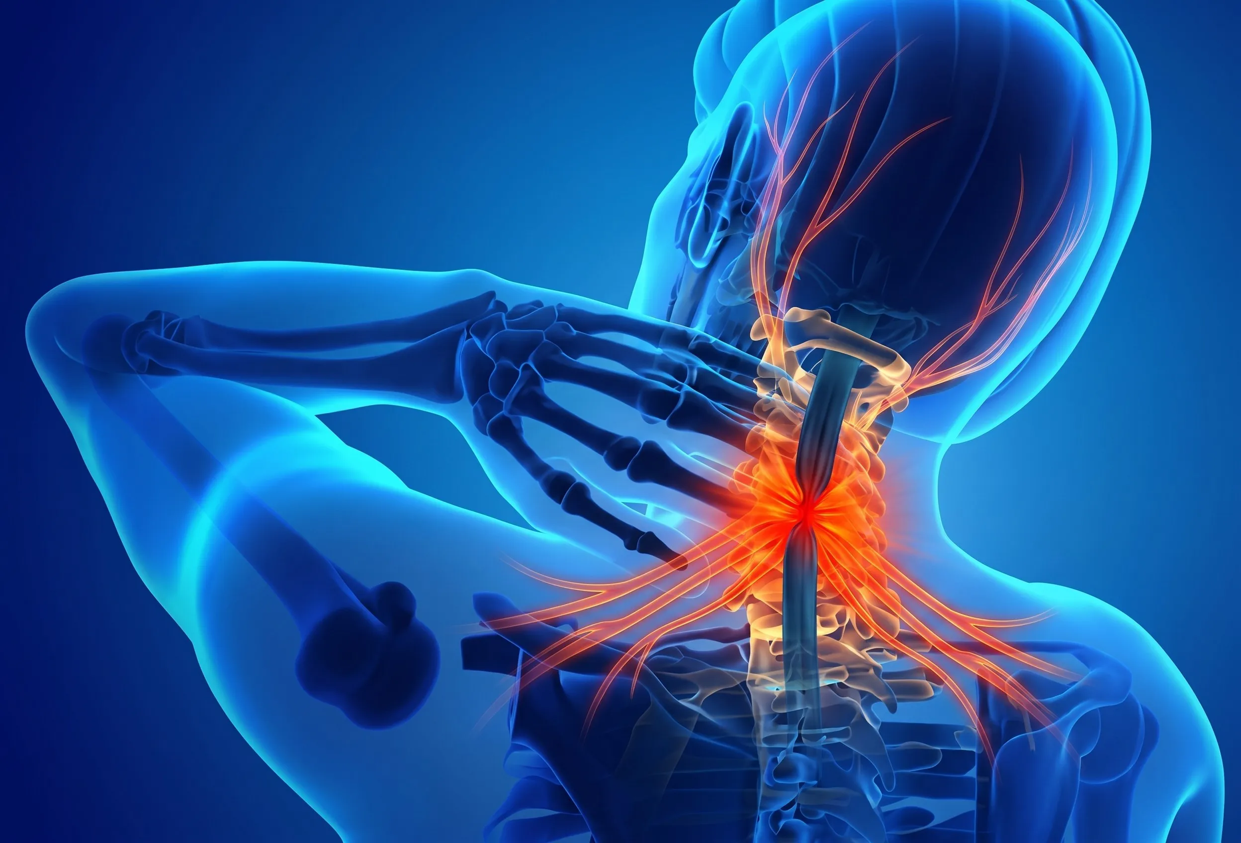
This narrowing leads to a hallmark clinical phenomenon known as Neurogenic Claudication:
- Standing & Walking Aggravation: When standing upright or walking, the lumbar spine naturally curves backward (Lordosis), further closing down the canal and choking blood supply to nerve roots, triggering heavy, cramped, and numb legs.
- The "Shopping Cart Sign": Sitting down or bending forward flexes the spine, expanding the spinal canal dimensions by 20% to 30%, which immediately restores blood flow and relieves leg fatigue.
Advanced Interventional Spine Care eliminates nerve swelling, washes away inflammatory adhesions, and precisely decompresses narrowed canals using stitchless keyhole endoscopy—freeing patients from pain without major open spinal fusion surgeries or titanium rods.
Types of Spinal Canal Stenosis
Understanding the precise anatomical zone of narrowing dictates the targeted non-surgical or keyhole approach.
1. Central Canal Stenosis
Narrowing of the main central spinal vault due to buckled ligamentum flavum and disc bulges, compressing the thecal sac and cauda equina.
- Symptoms: Bilateral leg heaviness, aching & fatigue while standing or walking.
- Classic Relief: Immediate comfort when bending forward or sitting down.
2. Lateral Recess Stenosis (Entrance Zone)
Narrowing of the gutter before the nerve enters the exit foramen, choked by hypertrophic superior articular facet spurs.
- Symptoms: Severe sharp radiating sciatica down one or both legs.
- Aggravated: Standing straight and walking uphill or downhill.
3. Foraminal Stenosis (Exit Zone)
Collapse of intervertebral disc height pinching the exiting spinal nerve root in its exit doorway (frequently at L5-S1 or L4-L5).
- Symptoms: Continuous burning calf, heel, or big toe numbness.
- Associated: Foot drop or weak push-off while walking.
Neurogenic Claudication vs. Vascular Claudication
Distinguishing nerve-compression claudication (Spinal Stenosis) from poor arterial blood circulation (Peripheral Artery Disease) is crucial for correct treatment.
| Clinical Feature | Neurogenic Claudication (Spine Stenosis) | Vascular Claudication (Blocked Arteries) |
|---|---|---|
| Underlying Cause | Nerve root compression in narrowed spinal canal | Insufficient arterial blood flow to leg muscles (PAD) |
| Relief Mechanism | Must sit down, squat, or bend forward (flex spine) | Relieved by standing still in place (no bending needed) |
| Stationary Bicycle Test | Painless (spine is flexed forward during cycling) | Painful (leg muscles demand oxygen regardless of posture) |
| Walking Downhill | Worse (downhill induces spine extension/narrowing) | Easier (requires less cardiovascular muscle exertion) |
| Pedal Pulses in Feet | Normal, strong, and palpable foot pulses | Diminished, weak, or absent foot pulses |
Our Diagnostic Protocol for Spinal Stenosis
Accurate clinical testing and cross-sectional imaging measure the exact reserve cross-sectional area of your spinal canal.
Claudication Distance
Treadmill walking distance tolerance vs. stationary bicycle test to confirm neurogenic origin of leg cramping.
Dynamic X-Rays
Standing flexion and extension views to detect degenerative spondylolisthesis (vertebra slipping forward over another).
3-Tesla MRI Spine
Measures thecal sac cross-sectional area (<75 mm² is severe), ligamentum flavum thickness, and lateral recess depth.
Diagnostic Block
Targeted selective nerve root block under live fluoroscopy to pinpoint the primary offending stenotic segment.
Non-Surgical & Keyhole Treatments for Stenosis
Minimally invasive daycare interventions performed under real-time fluoroscopic C-Arm guidance by Dr. Shrutika Bhagat in Patna.
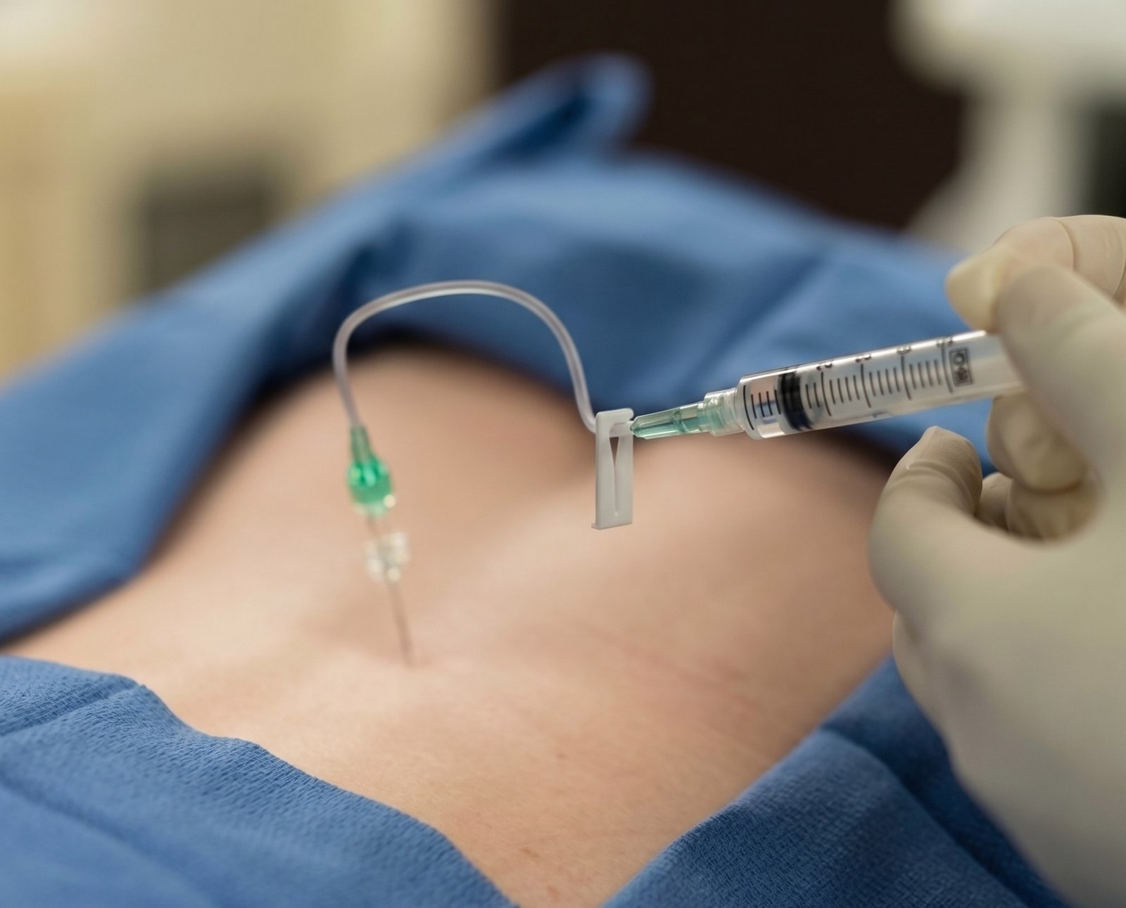 Image-Guided
Interlaminar & Caudal Epidurals
C-Arm fluoroscopy-guided multi-level wash delivering concentrated anti-inflammatory medication directly into the narrowed canal, shrinking swollen tissues and dramatically improving walking stamina.
- Rapidly relieves bilateral walking leg heaviness
- Restores functional spinal canal reserve volume
 Keyhole Surgery
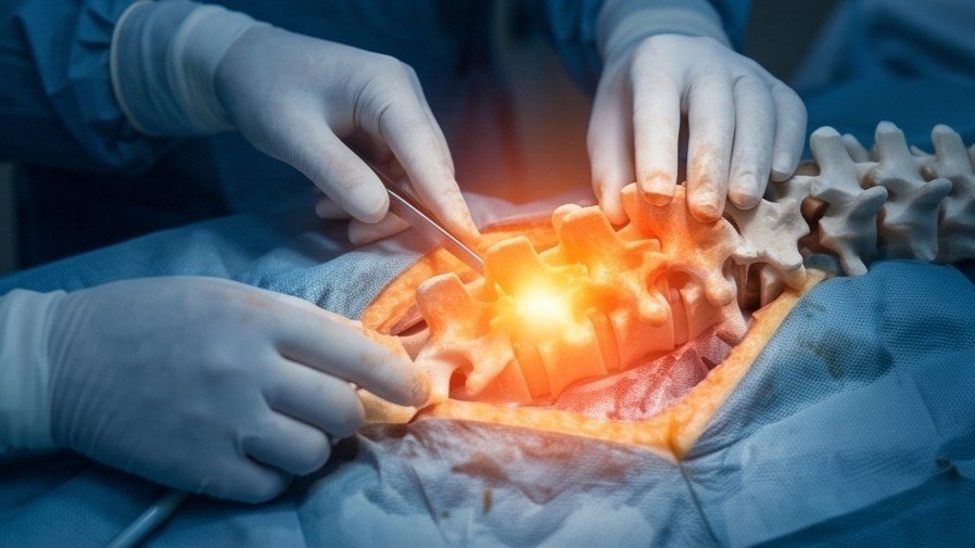
Keyhole Endoscopic Decompression
Ultra-minimally invasive stitchless decompression (TELD / IELD / UBE) through a tiny 7–8mm portal under local anesthesia. An HD camera visualizes the narrowed canal, precisely shaving thickened ligaments without spinal fusion.
- Zero metal rods, screws, or bone fusion
- Walk home within 2 hours of procedure
 Targeted Adhesiolysis
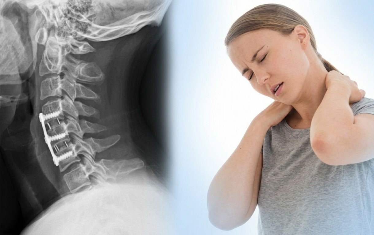
Epidural Catheter Neuroplasty
A specialized steerable micro-catheter (Racz catheter) navigates through the sacral hiatus under C-Arm guidance to gently dissolve dense fibrous adhesions and scar tissue choking the cauda equina.
- Mechanical release of entrapped cauda equina
- Highly effective for multi-level canal stenosis
 Pinpoint Accuracy
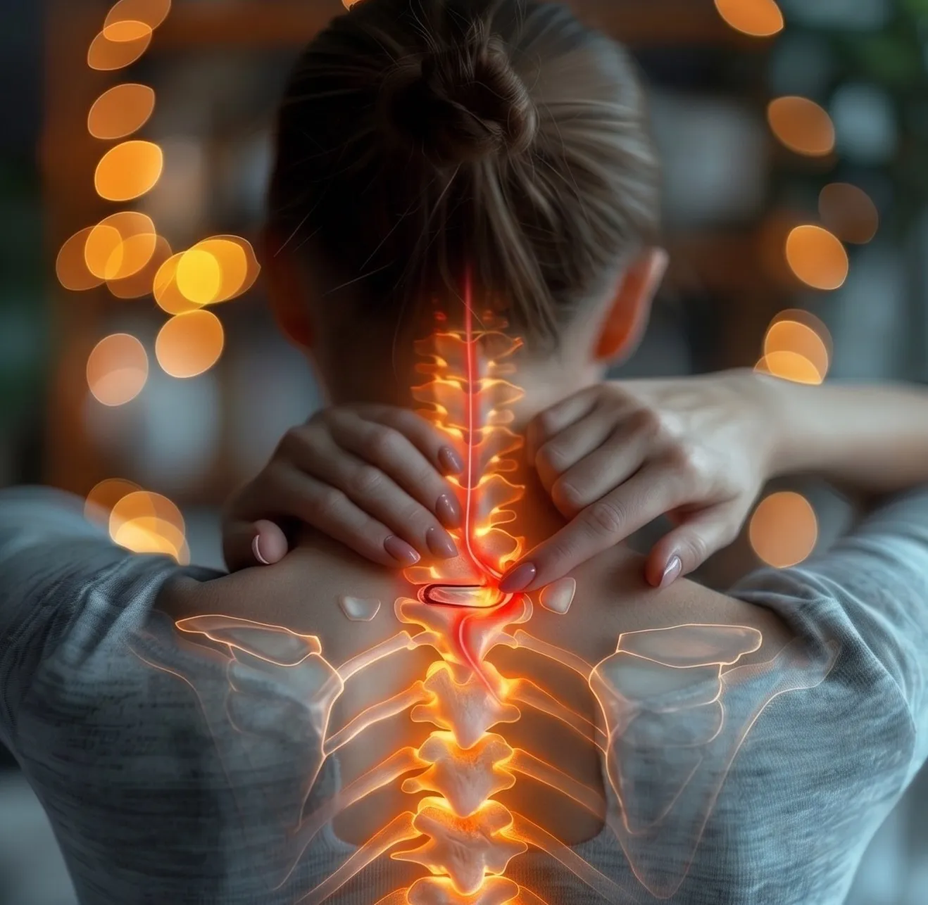
Targeted Transforaminal (TFESI)
Sub-millimeter fluoroscopy-guided injection directed at the lateral recess and exit neural foramen, relieving sharp unilateral radicular sciatica caused by foraminal bone spurs.
- Calms isolated nerve root pinches
- Stops shooting pain and foot tingling
 12-24 Months Relief
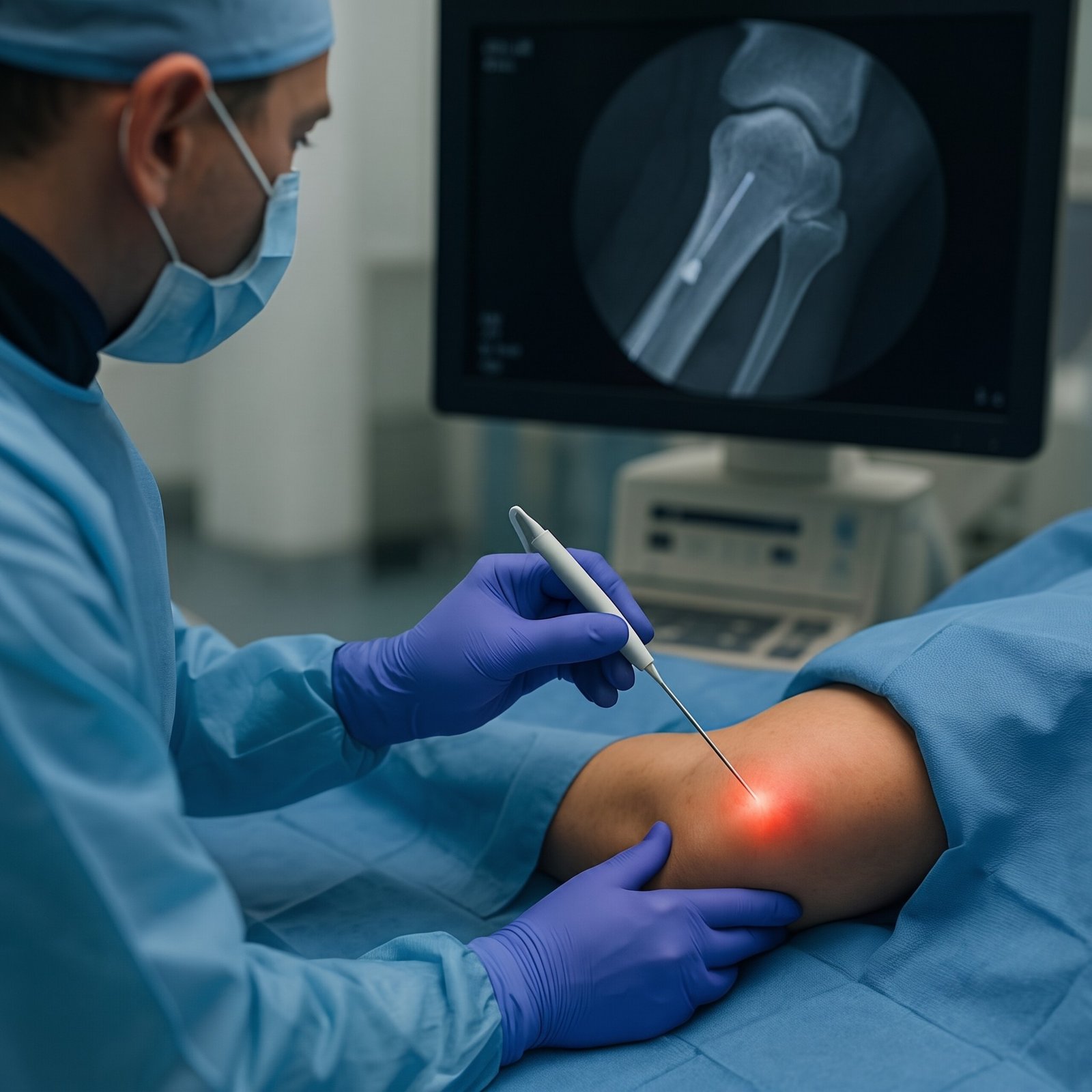
Facet Radiofrequency (RFA)
Thermal denervation of sensory medial branch nerves supplying enlarged, arthritic facet joints that crowd the spinal canal, eliminating accompanying severe lower back stiffness.
- Deactivates chronic arthritic facet pain
- Enhances standing upright tolerance
 Biologic Repair
Spine PRP & Ligament Biotherapy
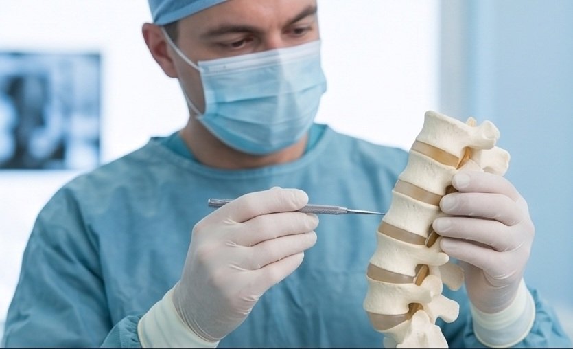
Autologous concentrated platelet-rich plasma (PRP) injected under live imaging to tighten lax spinal ligaments, preventing micro-instability and reducing compensatory ligamentous thickening.
- 100% natural healing growth factors
- Stabilizes degenerated lumbar motion segments
Open Laminectomy & Fusion vs. Keyhole Endoscopy
Why stitchless keyhole endoscopy and interventional procedures are transforming spinal stenosis care.
| Clinical Feature | Open Laminectomy & Fusion | Keyhole Endoscopic Decompression |
|---|---|---|
| Anesthesia Type | General Anesthesia (higher medical risk for elderly) | Local Anesthesia with conscious sedation (Safe for elderly) |
| Incision Size | Large 4 – 6 inch open surgical incision | Tiny 7–8mm keyhole port (Stitchless) |
| Bone & Muscle Resection | Extensive cutting of paraspinal muscles & lamina bones | Zero muscle cutting; preserves natural spinal stability |
| Metal Implants & Screws | Requires titanium screws, rods & cages | Zero metal implants or artificial hardware inserted |
| Hospital Stay | 4 to 7 days hospital admission | Daycare: Discharged within 2 to 4 hours |
| Recovery & Walking | 8 to 12 weeks of restricted bed rest & mobility | Immediate walking; resume routine life in 2–5 days |

Dr. Shrutika Bhagat
MBBS, MD, FIRA, FIAPM | Interventional Pain & Spine Endoscopy
Dr. Shrutika Bhagat brings super-specialized training in full-endoscopic spine surgery and image-guided interventional pain management, having successfully treated thousands of patients with severe back pain, disc prolapse, sciatica, nerve compression, and joint arthritis in Patna.
Spinal Canal Stenosis FAQs
Clear clinical answers regarding canal narrowing, walking claudication, epidural injections, and keyhole procedures.
Don't Let Chronic Pain Control Your Life.
Take the first step toward understanding your pain and exploring modern, evidence-based minimally invasive relief with Dr. Shrutika Bhagat.