Trigeminal Neuralgia Treatment in Patna
Permanent relief from excruciating electric-shock facial pain, jaw spasms, and chewing agony through precision C-Arm guided Gasserian Ganglion Radiofrequency Ablation without open brain surgery.
Understanding Trigeminal Neuralgia (The "Suicide Disease")
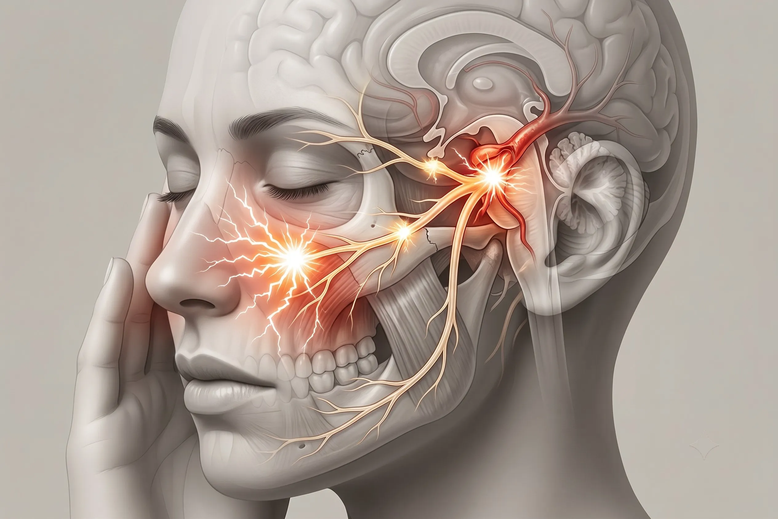
The Trigeminal Nerve (5th Cranial Nerve) is the primary sensory nerve supplying the entire face, teeth, gums, and jaw. It originates from the brainstem and branches out through a central nerve hub called the Gasserian (Trigeminal) Ganglion into three distinct pathways:
- V1 (Ophthalmic): Forehead, eyes, and top of the nose.
- V2 (Maxillary): Cheek, upper lip, upper teeth, and roof of the mouth.
- V3 (Mandibular): Lower jaw, lower teeth, chin, and side of the face.
Trigeminal Neuralgia (Tic Douloureux) occurs when a pulsating blood vessel (such as the Superior Cerebellar Artery) compresses and strips the protective myelin insulation off the nerve root. This causes hyper-synchronized cross-talk: trivial touch signals (light breeze, brushing teeth, chewing food, or talking) detonate blinding paroxysms of electric-shock lightning bolts across one side of the face.
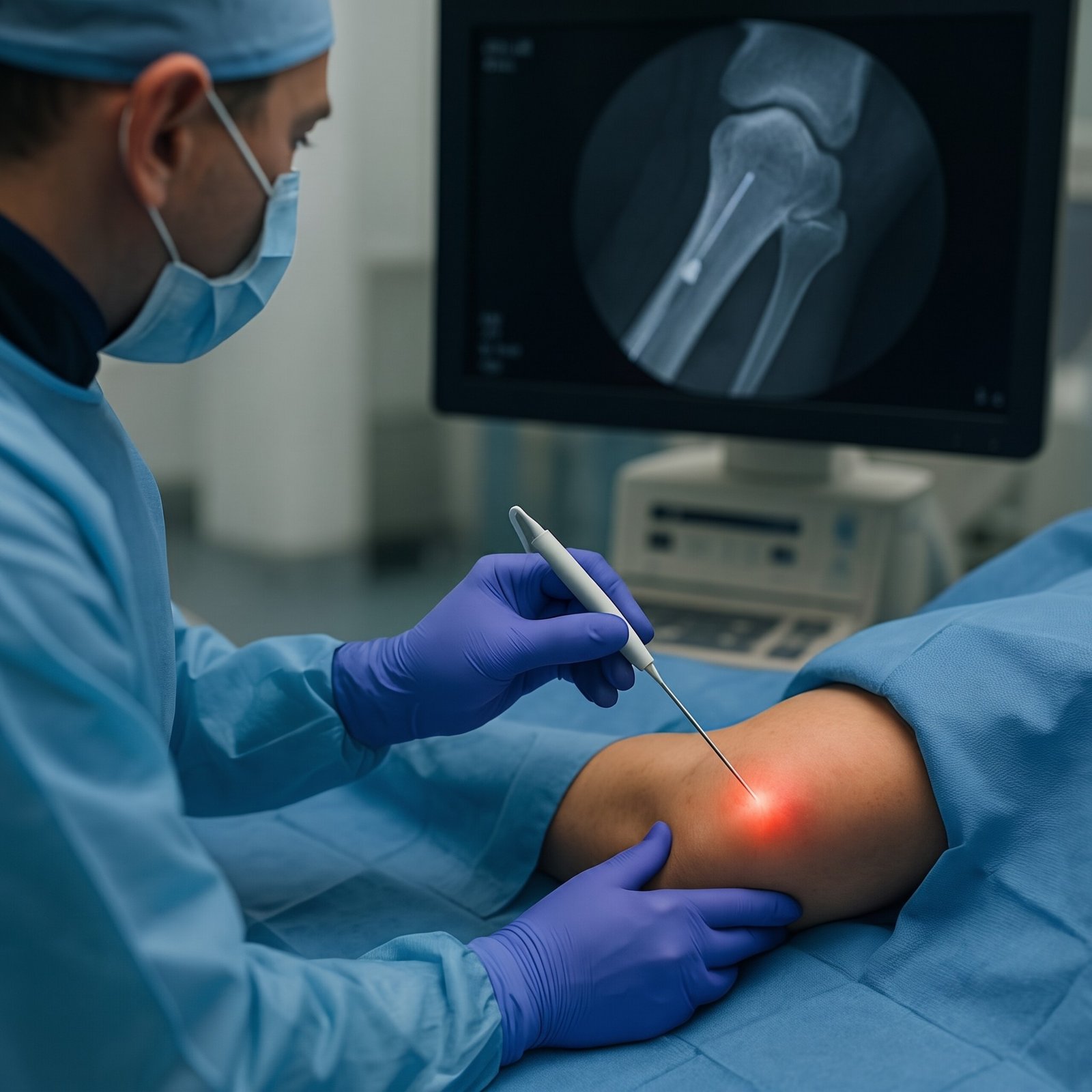
Gasserian Ganglion Radiofrequency Ablation (RFA) provides 2 to 5 years of profound, immediate pain relief by percutaneously deactivating the pain-transmitting sensory fibers inside the foramen ovale under live C-Arm guidance—restoring normal eating and smiling in a 20-minute daycare procedure without opening the skull.
Clinical Presentation of Trigeminal Neuralgia
Differentiating trigeminal neuralgia from dental toothaches, TMJ dysfunction, and atypical facial pain.
Sudden Electric-Shock Paroxysms
Intense, knife-like, lightning-bolt shooting pain lasting from a fraction of a second up to 2 minutes, strictly confined to one side of the face.
Innocuous Touch Triggers
Triggered by non-painful stimuli: brushing teeth, shaving, applying makeup, washing the face with water, smiling, chewing, or even a light cold breeze.
Unnecessary Tooth Extractions
Frequently misdiagnosed as dental abscesses. Many patients undergo multiple root canals and extractions with zero relief before seeing a pain specialist.
Our 4-Step Diagnostic Protocol for Facial Pain
Pinpointing the exact trigeminal nerve division and ruling out secondary brain tumors or multiple sclerosis.
Trigger Zone Mapping
Detailed clinical mapping of V1, V2, and V3 dermatomes to identify trigger zones and corneal reflex integrity.
3D CISS/FIESTA MRI
Gold Standard: High-resolution brainstem MRI sequences visualizing neurovascular conflict between artery and nerve root.
Multiple Sclerosis Screen
Screening brain MRI for demyelinating brainstem plaques in young patients presenting with bilateral facial pain.
Diagnostic Branch Block
Ultrasound/Fluoroscopy test block of peripheral branch. Immediate elimination of trigger pain confirms RFA candidacy.
Non-Surgical Interventions for Trigeminal Neuralgia
Pinpoint daycare procedures performed under live fluoroscopic C-Arm guidance by Dr. Shrutika Bhagat in Patna.
 Gold Standard
Gasserian Ganglion RFA (Hartel's Approach)
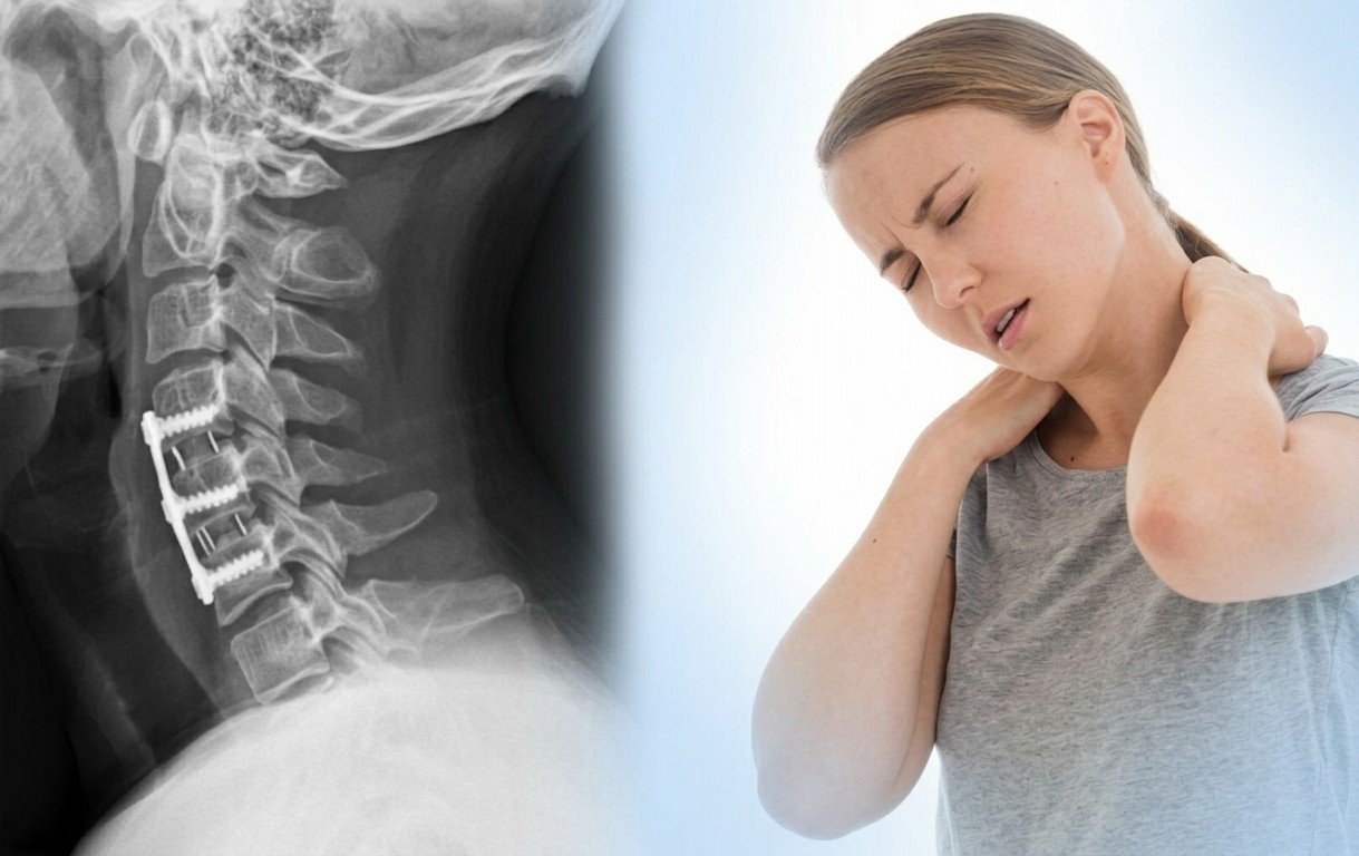
Under live C-Arm fluoroscopy, a radiofrequency cannula enters through the cheek into the foramen ovale. Precision thermal neurolysis selectively coagulates pain-carrying A-delta and C fibers while preserving motor jaw function.
- Provides 2 to 5 years of freedom from pain
- 20-minute daycare procedure, zero craniotomy
 Corneal Safe
Pulsed Radiofrequency (PRF) Neuromodulation
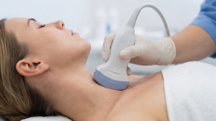
Non-destructive electromagnetic pulses applied to upper trigeminal branches (V1 and V2) to reset hyper-excited nerve transmission without causing facial numbness or risking corneal sensation loss.
- 100% safe for forehead and eye branch pain
- Preserves normal facial tactile sensation
 Targeted Blocks
Peripheral Branch Blocks & Neurolysis
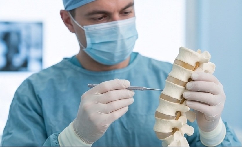
Direct ultrasound-guided blocks of the supraorbital, infraorbital, or mental nerves using local anesthetic and neurolytic agents to immediately stop severe acute breakthrough pain paroxysms.
- Fast emergency relief in acute crisis
- Outpatient clinic procedure in 5 minutes
 Mechanical Compression
Balloon Microcompression
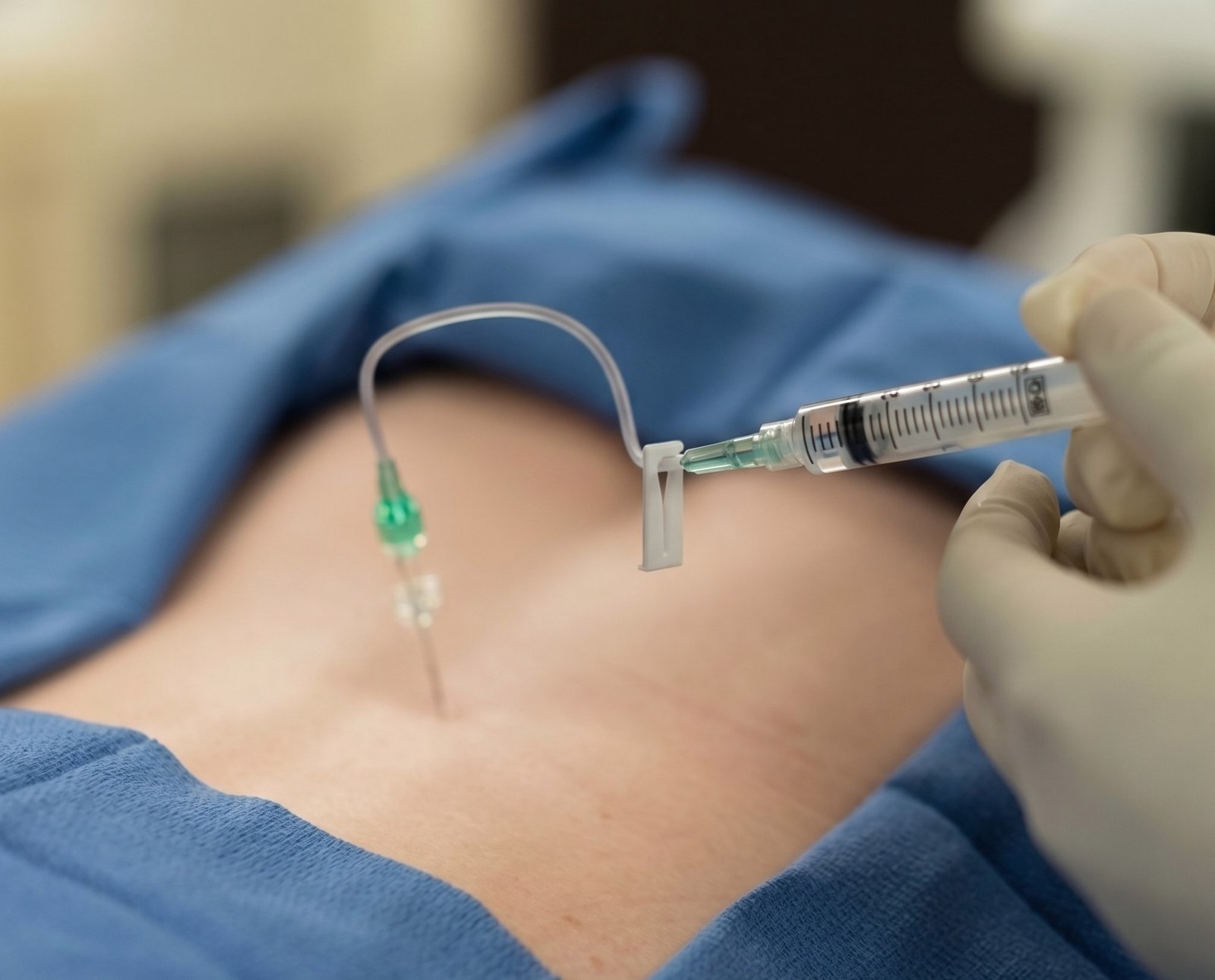
A tiny Fogarty balloon catheter is inflated inside Meckel's cave under fluoroscopy to mechanically compress the Gasserian ganglion, ideal for refractory V1 involvement and elderly patients.
- Excellent alternative for upper-face pain
- High immediate success rate
 Facial Hydrodissection
Sphenopalatine & Maxillary Hydrodissection
Ultrasound/fluoroscopy delivery of fluid to release entrapped maxillary nerve fibers in the pterygopalatine fossa, suppressing deep facial burning and cluster headache overlaps.
- Calms deep cheek and sinus burning
- Discharges sympathetic facial tension
 Medical Optimization
Precision Drug Titration
Scientific titration of voltage-gated sodium channel blockers (Carbamazepine, Oxcarbazepine, Baclofen, Lamotrigine) to achieve maximum pain suppression with minimal side effects.
- Eliminates heavy drug drowsiness
- Guided tapering after successful RFA
Open Brain Surgery (MVD) vs. Daycare Gasserian RFA
Why Gasserian Ganglion Radiofrequency Ablation is the safest, most effective first-line interventional standard.
| Clinical Feature | Open Brain Surgery (Microvascular Decompression - MVD) | Daycare Gasserian Ganglion RFA |
|---|---|---|
| Anesthesia Required | General Anesthesia (High risk for elderly & cardiac patients) | Local Anesthesia + Light Sedation (Extremely safe for all ages) |
| Incision & Skull Opening | Craniotomy (Surgical hole drilled behind the ear to open skull) | Zero incisions (Pinhole needle entry through cheek) |
| Brainstem & Hearing Risks | Risk of stroke, meningitis, CSF leakage & permanent hearing loss | Zero brain retraction (Purely extracranial percutaneous route) |
| Hospital Stay | 5 to 8 days in ICU and neurosurgical ward | Daycare: Discharged home within 2 to 3 hours |
| Repeatability | Re-exploration craniotomy is technically hazardous | Easily repeatable if pain ever recurs years later |

Dr. Shrutika Bhagat
MBBS, MD, FIRA, FIAPM | Interventional Pain & Spine Endoscopy
Dr. Shrutika Bhagat brings super-specialized training in full-endoscopic spine surgery and image-guided interventional pain management, having successfully treated thousands of patients with severe back pain, disc prolapse, sciatica, nerve compression, and joint arthritis in Patna.
Trigeminal Neuralgia FAQs
Clinical answers regarding Gasserian RFA, avoiding brain surgery, medication side effects, and permanent relief.
Don't Let Chronic Pain Control Your Life.
Take the first step toward understanding your pain and exploring modern, evidence-based minimally invasive relief with Dr. Shrutika Bhagat.