Vertebral Fracture Treatment in Patna
Rapid same-day relief from excruciating osteoporotic back pain & spine collapse through precision daycare Percutaneous Vertebroplasty and Kyphoplasty.
Understanding Osteoporotic Vertebral Compression Fractures (VCF)
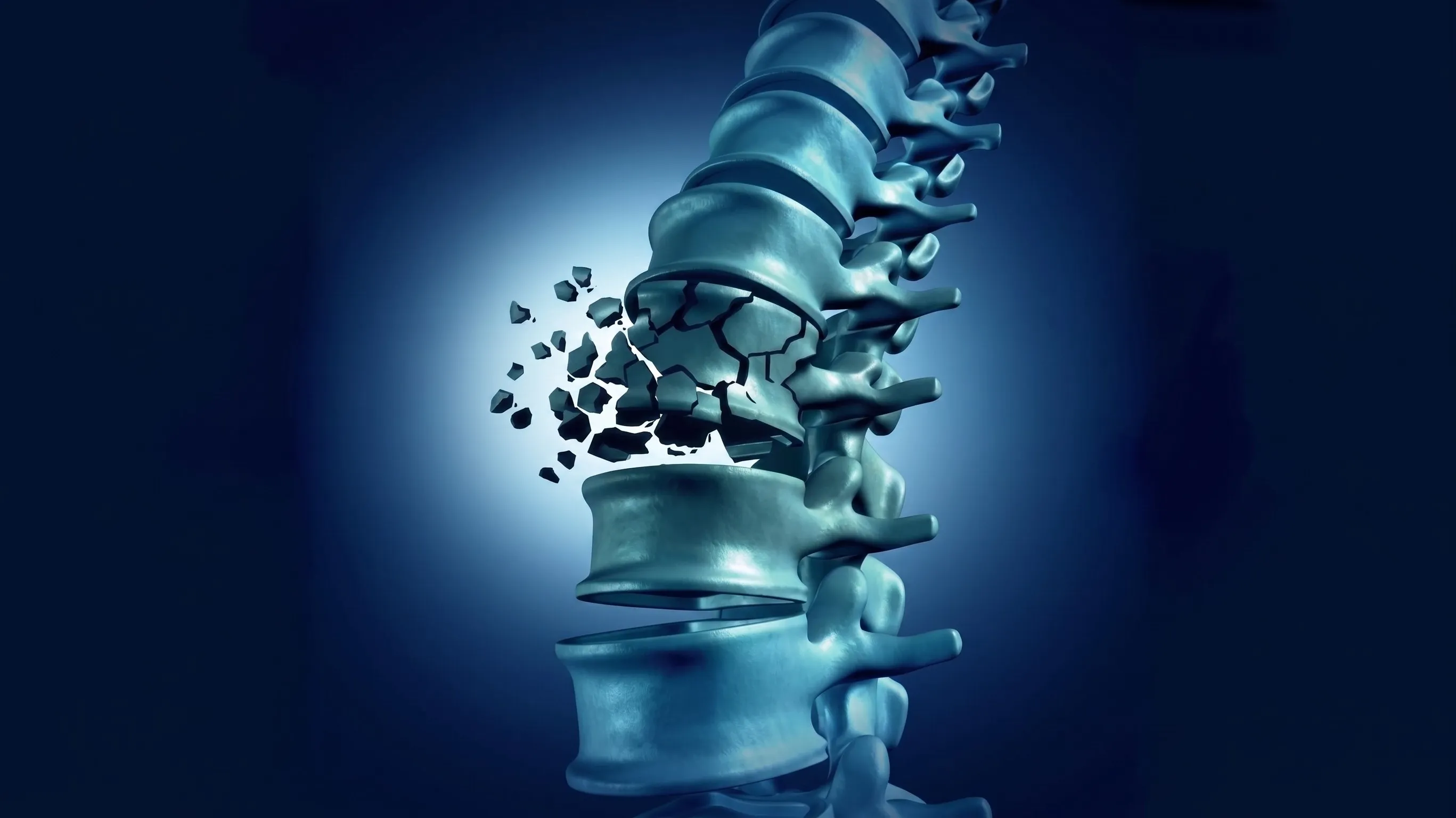
A Vertebral Compression Fracture (VCF) occurs when the sponge-like trabecular bone inside a spinal vertebral body collapses under load. In individuals with Osteoporosis (porous, brittle bones) or osteopenia, even trivial everyday actions—such as coughing, sneezing, bending to pick up an object, or stepping off a low curb—can cause the bone to buckle and fracture.
Unlike long bone fractures in the arm or leg, a fractured spinal vertebra cannot be immobilized with a simple plaster cast. Every breath, cough, and slight movement causes Micro-Motion at the fracture site, firing intense pain signals from the densely innervated periosteal nerve endings.
If left untreated, vertebral compression fractures lead to:
- Severe Midline Back Pain: Inability to turn in bed, sit upright, or stand without agonizing distress.
- Progressive Spinal Kyphosis ("Dowager's Hump"): Forward stooping posture that crowds abdominal organs and compromises lung capacity.
- Cascade of Future Fractures: Altered spinal biomechanics shifting heavy stress onto adjoining vertebrae, multiplying fracture risk.
Percutaneous Vertebroplasty / Kyphoplasty provides immediate, transformative relief by injecting high-strength medical bone cement through a needle under live C-Arm imaging—instantly solidifying the bone, eliminating micro-motion, and restoring mobility on the very same day.
Types & Patterns of Vertebral Fractures
Accurate classification on high-resolution MRI and X-ray determines whether Vertebroplasty or Kyphoplasty is the ideal solution.
Wedge Compression Fracture
The front (anterior) wall of the vertebral body collapses while the posterior wall remains intact, producing a characteristic wedge shape that causes forward stooping.
- Symptoms: Severe midline backache, height loss, stooping.
Biconcave (Codfish) Fracture
The central portion of the vertebral endplates sinks inward under disc pressure while the peripheral cortex remains relatively intact.
- Symptoms: Deep dull aching, severe stiffness when rolling in bed.
Crush / Total Collapse
The entire vertebral body (anterior, middle, and posterior columns) collapses uniformly, causing significant vertical height loss and acute spinal instability.
- Symptoms: Complete inability to walk or stand, severe spasms.
Pathological / Malignant Fracture
Vertebral collapse caused by metastatic lesion, multiple myeloma, or severe steroid-induced osteoporosis weakening the vertebral architecture.
- Symptoms: Constant deep nocturnal aching and severe bone agony.
Where Do Vertebral Fractures Strike Most?
Over 90% of osteoporotic compression fractures occur at the Thoracolumbar Junction, where the rigid ribcage transitions into the flexible lumbar spine.
Thoracolumbar Junction (D11, D12, L1, L2)
The mechanical pivot point between the stable thoracic cage and mobile lumbar column. D12 and L1 sustain the highest stress concentrations during bending and coughing.
Acute intense pain across the mid-back and flank, radiating around the lower ribcage and groin. Severe pain upon turning in bed.
Lower back agony preventing weight-bearing, standing, or sitting. Severe paraspinal muscle spasms locking the lower spine.
Mid-Thoracic Spine (D6, D7, D8)
Common in advanced osteoporosis. Multiple sequential wedge collapses here cause the characteristic round-shouldered hunchback posture (Kyphosis).
Sharp knife-like pain between the shoulder blades, worsening on deep inspiration, coughing, or laughing.
Progressive height loss (2 to 5 inches), rib cage crowding against pelvic bones, and shortness of breath.
Our Diagnostic Protocol for Spine Fractures
Distinguishing a fresh, acute painful fracture from an old healed collapse is essential to determine whether Vertebroplasty will provide relief.
Spinous Percussion
Gentle palpation over the spinal spinous processes precisely pinpoints the exact acute fractured vertebra.
Digital Spine X-Rays
Standing AP and lateral views quantify vertebral height loss, wedge percentage, and the Cobb angle of kyphotic curvature.
3T MRI (STIR Sequence)
Gold Standard: Bright bone marrow edema on STIR sequences confirms a fresh, active fracture responsive to cement.
DEXA Bone Scan
Measures Bone Mineral Density (BMD T-score) to classify osteoporosis severity and guide targeted anti-osteoporotic drug therapy.
Minimally Invasive Interventions for Spine Fractures
Daycare pinhole procedures performed under live fluoroscopic C-Arm guidance by Dr. Shrutika Bhagat in Patna.
 Gold Standard
Percutaneous Vertebroplasty
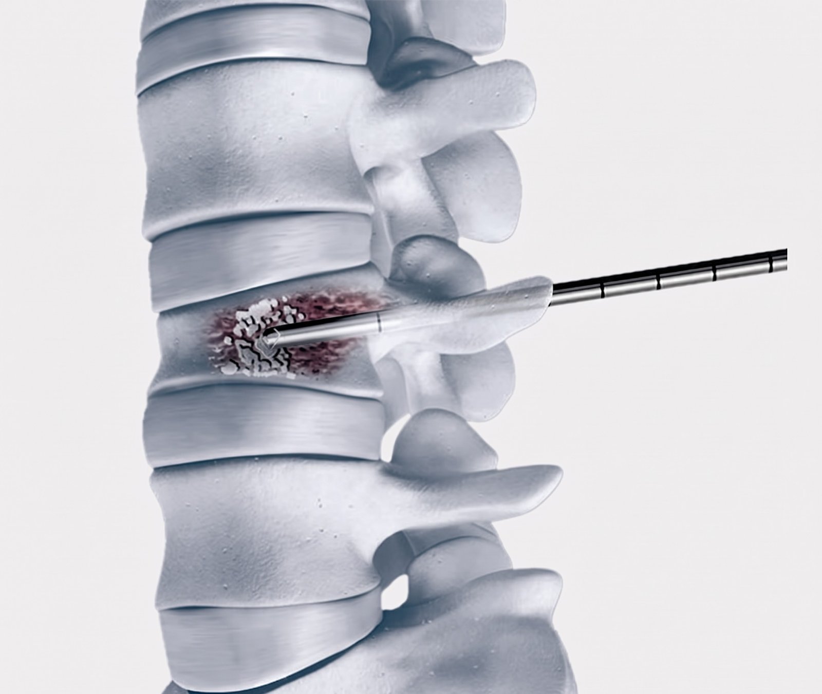
Under live C-Arm fluoroscopy and local anesthesia, a fine 11-13G needle is guided through the pedicle into the fractured vertebra. Medical-grade bone cement (PMMA) is injected, instantly solidifying the collapse and eliminating micro-motion pain within minutes.
- Pain relief within 1 hour; walk same day
- 2mm pinhole puncture; zero surgical stitches
Height Restoration
Balloon Kyphoplasty
An inflatable orthopedic balloon is gently expanded inside the collapsed vertebral body to restore lost vertebral height and correct kyphotic deformity. The balloon is deflated, leaving a cavity filled with thick bone cement at low pressure.
- Restores natural spinal height & posture
- Ultra-low risk of cement leakage
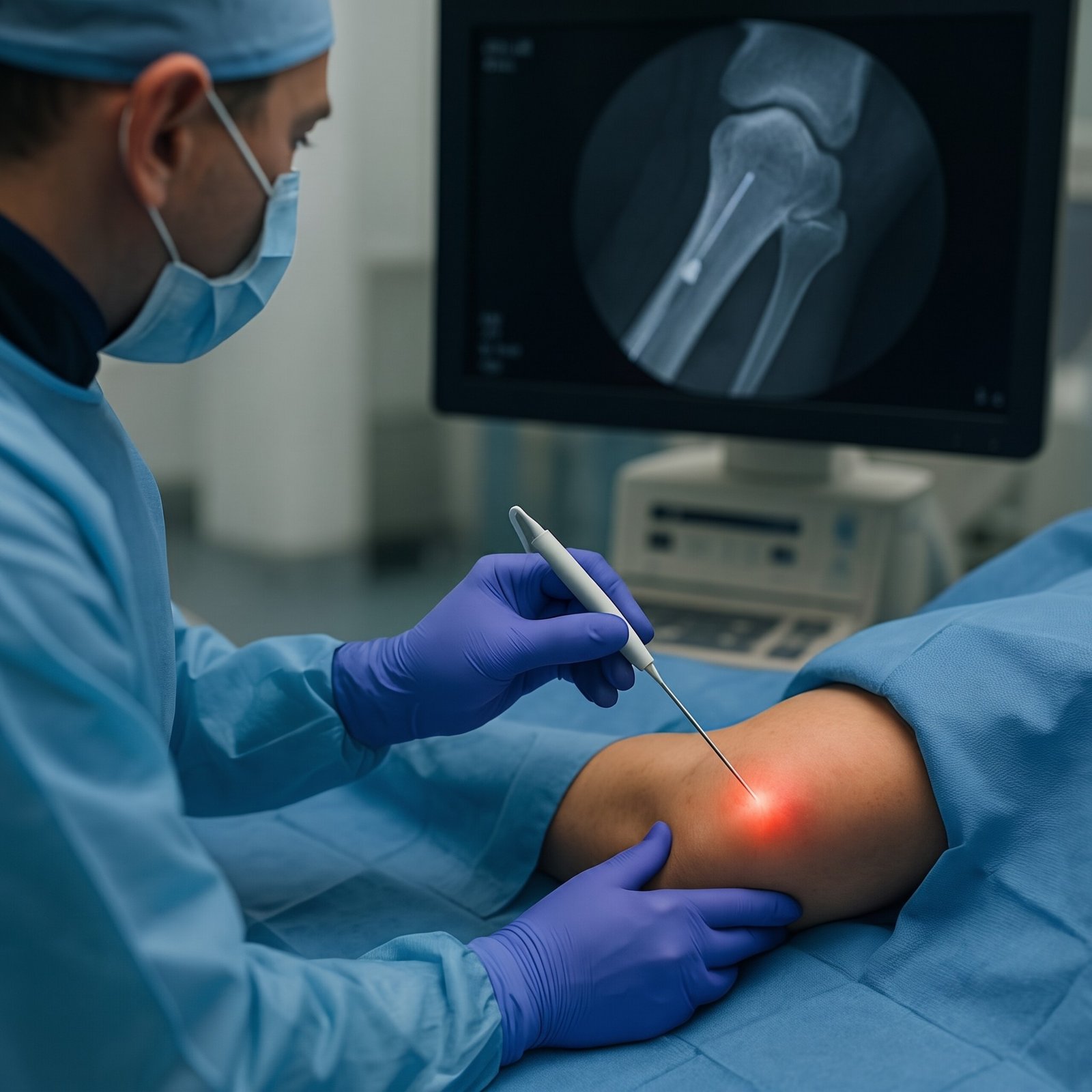 Instant Spasm Relief
Paraspinal Nerve Blocks
Precision image-guided local anesthetic and anti-inflammatory injection targeting the dorsal rami and medial branches at the fractured level, instantly interrupting severe reflexive paraspinal muscle spasms.
- Fast relief from locking back spasms
- Enables comfortable sitting and mobility
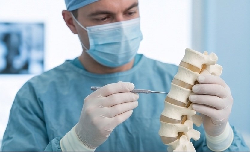 Bone Strengthening
Anabolic Bone Building Therapy
Comprehensive medical management with bone-forming agents (Teriparatide) or antiresorptives (Zoledronic Acid / Denosumab) to rebuild trabecular bone density and prevent future fractures.
- Stops future adjacent vertebral fractures
- Strengthens entire skeletal framework
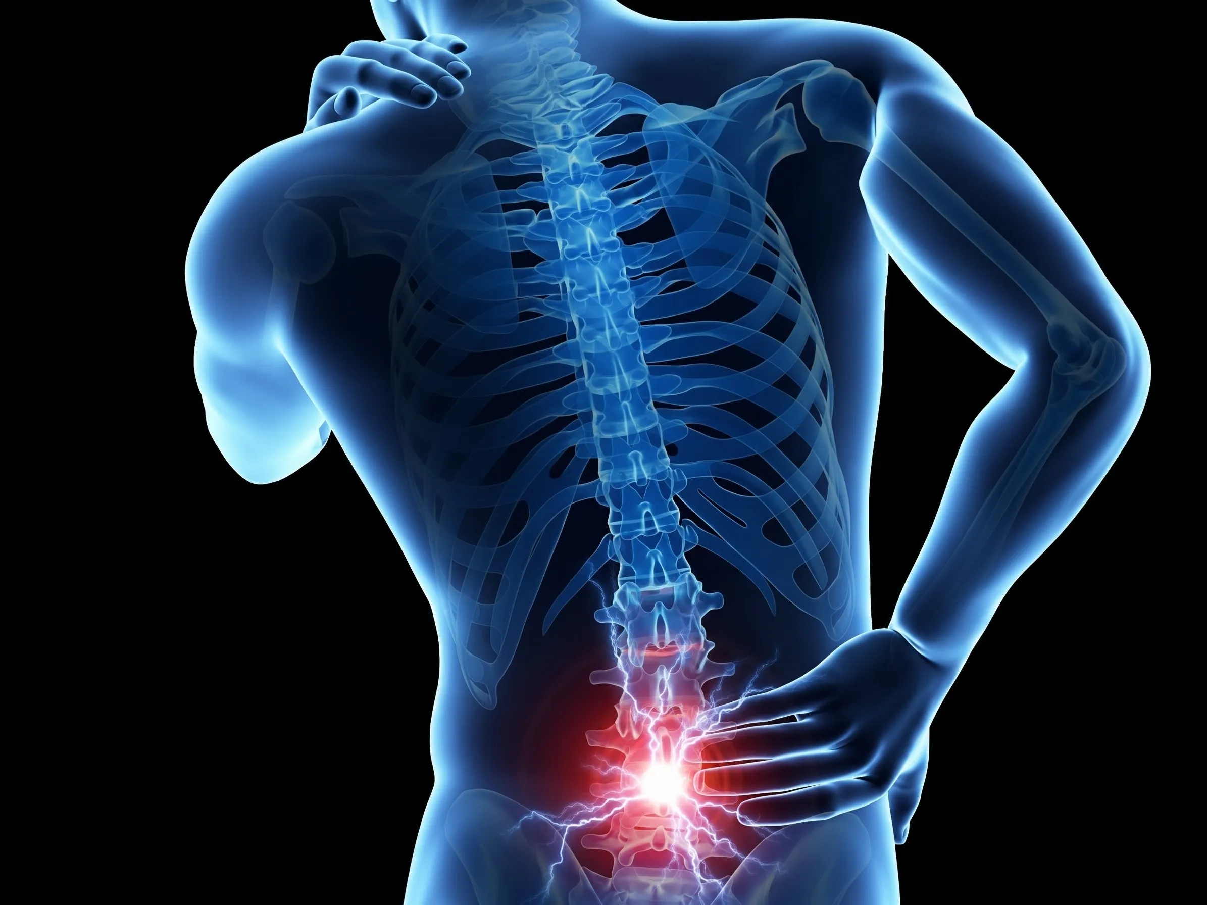 External Support
Custom Thoraco-Lumbar Bracing
Custom-molded lightweight Taylor / CASH hyperextension braces designed to offload anterior vertebral column pressure during the acute consolidation phase, preventing progressive kyphosis.
- Offloads stress from fractured bone
- Prevents forward postural collapse
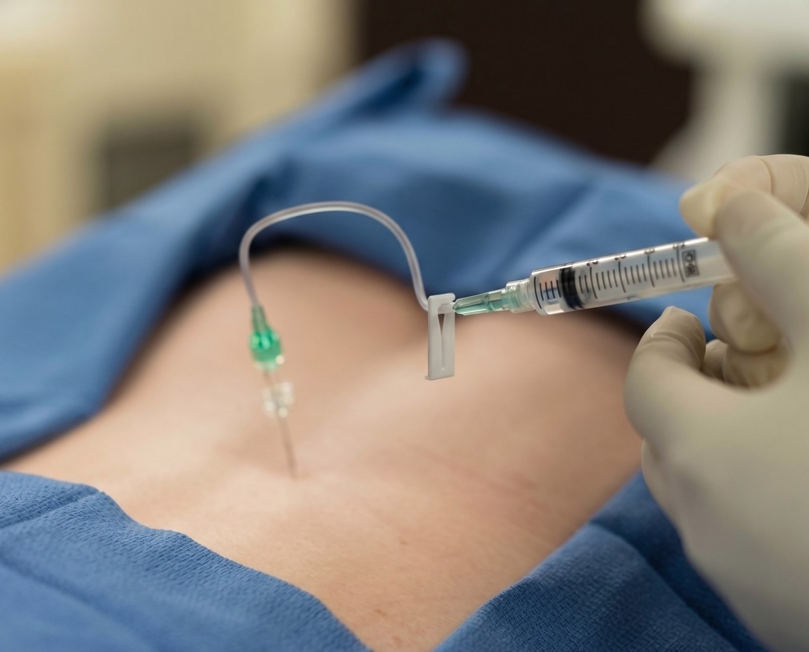 Biologic Repair
Regenerative Ligament PRP
Autologous concentrated platelet-rich plasma (PRP) injected under ultrasound into strained interspinous ligaments and facet capsules to promote biological tissue repair and resolve chronic secondary ligamentous pain.
- 100% natural, patient's own growth factors
- Repairs stretched paraspinal ligaments
Open Spine Surgery vs. Percutaneous Vertebroplasty
Why minimally invasive bone cement stabilization is the treatment of choice for osteoporotic spine fractures.
| Clinical Feature | Open Spine Surgery & Pedicle Screws | Percutaneous Vertebroplasty / Kyphoplasty |
|---|---|---|
| Anesthesia Required | General Anesthesia (Dangerous in frail elderly) | Local Anesthesia + Mild Sedation (Extremely safe) |
| Incision & Stitches | Large 4 – 6 inch incision with heavy blood loss | Tiny 2mm needle puncture (Zero stitches) |
| Screw Loosening in Brittle Bone | High failure rate (Screws pull out of soft osteoporotic bone) | Cement anchors directly into brittle trabeculae (Zero screw risk) |
| Time to Pain Relief | Weeks to months of post-operative surgical pain | Immediate pain relief within 30 to 60 minutes |
| Hospital Stay & Walking | 5 to 10 days admission; weeks of bed rest | Daycare: Discharged in 2 to 3 hours; walking same day |

Dr. Shrutika Bhagat
MBBS, MD, FIRA, FIAPM | Interventional Pain & Spine Endoscopy
Dr. Shrutika Bhagat brings super-specialized training in full-endoscopic spine surgery and image-guided interventional pain management, having successfully treated thousands of patients with severe back pain, disc prolapse, sciatica, nerve compression, and joint arthritis in Patna.
Vertebral Fracture FAQs
Clear clinical answers regarding Vertebroplasty, bone cement safety, recovery timeline, and osteoporosis prevention.
Don't Let Chronic Pain Control Your Life.
Take the first step toward understanding your pain and exploring modern, evidence-based minimally invasive relief with Dr. Shrutika Bhagat.